


INTRODUCTION
More than 180 000 hip and knee joint replacements are performed annually in the UK,1 and this number will rise with an increasing ageing population. Hip and knee replacements are largely considered successful in terms of patient-reported outcome measures, survivorship, and low complication rates. However, even a 1% average infection rate after prosthetic replacement represents an increasing burden to the NHS.2
Infected prostheses can present early or late3 and the treatment for each differs. As a general rule the acute (early) infections are much more clinically apparent than the ‘never been right’ chronic infection.
DIAGNOSIS
Early infections
Defined as occurring less than 3 months post-surgery,3 these tend to present with cellulitis and symptoms of sepsis. The wound may have discharged since the time of surgery or after the wound had healed initially. A persistent serous, haemo-serous, or purulent discharge may indicate infection. The knee is tender and swollen, with evidence of inflammation. As swelling, tenderness, pain, and minor serous wound discharge can be normal early after knee replacement, this may pose a diagnostic challenge in differentiating normal from infected.
Late infections
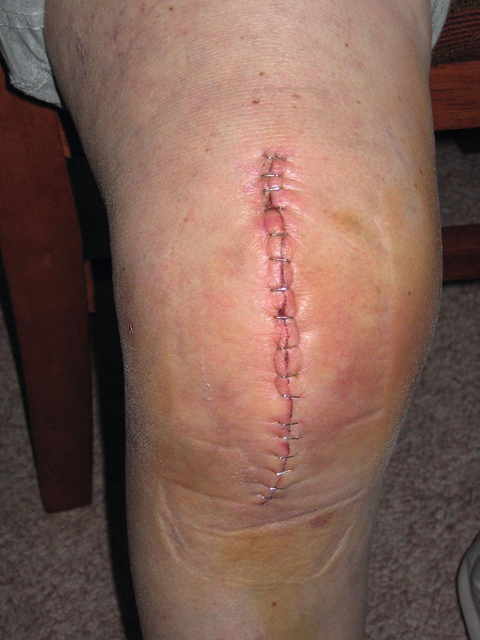
Defined as occurring more than 3 months post-surgery,3 late infections present more insidiously (Figure 1). The patients often report the knee has ‘never been right’ or that their preoperative arthritic pain was never eradicated. They may have had problems of wound discharge after surgery. They may even have had courses of antibiotics in the postoperative period. They frequently, however, have no useful clinical signs to elicit; the knee can be non-swollen, move well, have no erythema, and be non-tender on palpation even though ongoing pain is the presenting complaint. The blood tests and X-rays are frequently normal.

Flowchart of management of suspected prosthetic joint infection using total knee replacement (TKR) as model.

Slightly pink scar. Unlikely to represent infection. Source: Ravedave, Wikimedia Commons. This file is licensed under the Creative Commons Attribution-Share Alike 2.5 Generic, 2.0 Generic, and 1.0 Generic licence.
INVESTIGATIONS
In a full blood count (FBC) test, white cell count may be elevated or normal. C-reactive protein (CRP) is invariably elevated (>10 mg/ml in mild and >100 mg/ml in more severe) in acute infection. CRP can however be normal in chronic infection.
X-rays may be normal in early and late infections, but chronic infection is capable of causing prosthesis loosening and bone destruction. A bone scan can demonstrate an infected prosthesis in chronic infections. A negative bone scan, however, does not exclude a prosthetic infection. Joint fluid aspiration is a highly sensitive test for infection and has the added benefit of also growing the organism concerned with its antibiotic sensitivity and resistance profile. Prosthetic joints should only be aspirated in the setting of a sterile operating theatre.

Grossly cellulitic knee with swelling. Definitely infected. Copyright Matthew S Austin, MD. Used with permission. Originally published in Diaz-Ledezma C, Austin MS. Markers for diagnosing a periprosthetic knee infection, on ICJR.net, the website of the International Congress for Joint Reconstruction. http://icjr.net/article_92_knee_pji.htm#.Va1hyUVrXwM (accessed 12 Jan 2017).
MANAGEMENT OF EARLY PRESENTING PROSTHETIC INFECTION
The overriding principle in early infections is to attempt infection eradication while preserving the implant. This ‘implant preservation’ philosophy is done via a technique known as ‘DAIR’ meaning debridement, antibiotics, and implant retention.4 This entails not starting antibiotics until samples are taken under sterile conditions at the time of operation, to maximise the chance of culturing the infective organism, and tailoring antibiotic treatment, an open debridement (meaning sharp dissection of the infected soft tissues), and washout with sterile saline under pressure-pulsed lavage. This treats the protective ‘membrane’ (glycocalyx gel, which confers a protection from antibiotics) that forms over the bacteria, while the bacteria are on the prostheses. If this membrane is not removed, treatment failure frequently occurs.5 Broad-spectrum antibiotics are started immediately after taking the culture samples, and are continued intravenously (IV) until an organism is identified and a more directed antibiotic treatment can be given. Antibiotics are continued IV for 6 weeks, then converted to oral agents, typically for another 6 weeks in hip infections and longer in knee infections. The success rate of DAIR with retention of the prostheses in early infections is around 80%.4
The evidence supports early intervention with surgery and only then antibiotics (long- term IV then oral), rather than treatment with a short course of antibiotics in the community. Community antibiotics may only delay or complicate diagnosis,6 and much more importantly delay treatment as well as possibly conferring antibiotic resistance to the original infective organism (particularly relevant considering already increasing antibiotic resistance and its consequences).
MANAGEMENT OF LATE PRESENTING PROSTHETIC INFECTION
Successful implant retention reduces significantly beyond 3 months post-implantation.6 Late presenting infections are most successfully treated with removal of the implants either in a single-stage procedure or in two stages. Either way involves thorough debridement and washout, and the use of IV antibiotics.
Reasons for the high failure rate of implant retention in late presenting infection include the depth of infection in the bone by this stage, development of a mature membrane, and multiple infecting organisms.6
TREATMENT ALGORITHM
Best practice suggests that patients presenting to their GP with suspicion of prosthetic joint infection should not be started on a short course of oral antibiotics. This risks delaying diagnosis, delaying treatment, reducing the chance of culturing the organism at time of washout surgery, and conferring antibiotic resistance to the original infecting organism.
The patient should, in the case of suspected early infection, be referred to the on-call orthopaedic team who would then investigate and start definitive treatment.
In the case of late presenting infection, this represents a non-urgent problem, and best practice involves an outpatient referral to an orthopaedic surgeon (preferably the original operating surgeon).
The difficult problem of the acutely presenting knee after recent surgery with bruising, swelling, and a slightly pink wound in a slightly warm knee represents diagnostic difficulty. This should be referred to the orthopaedic on-call team, and there may be a place for the patient being seen in the following morning’s fracture clinic (in systems that are set up with slots for this purpose) in cases where the patient is definitely not septic, but there is doubt as to the diagnosis of infection. The decision to operate, including aspiration of the joint in a sterile operating theatre environment, is a complex one.
Notes
Provenance
Freely submitted; externally peer reviewed.
Competing interests
The authors have declared no competing interests.
Discuss this article
Contribute and read comments about this article: bjgp.org/letters
- Received February 22, 2016.
- Revision requested February 25, 2016.
- Accepted July 25, 2016.
- © British Journal of General Practice 2017
In this issue


Jump to section
More in this TOC Section
Related Articles
Cited By...

