


Photography has been used extensively in medicine since the 19th century, and now the universal availability of the digital camera has made the recording of images, archiving, and their transmission over the internet straightforward. When downloaded onto the practice computer, pristine images can, at the touch of a few keys, appear and can be used not only as accurate visual records, but can be sent anywhere for specialist advice regarding diagnosis and treatment. The common adoption of computerised archives should have encouraged a greater use of simple digital imaging, but written text continues to dominate our records. Our patients, themselves, store and transmit vast numbers of digital images through the internet, but we are slow in keeping up with this changed social environment.
Although GPs often send their interesting clinical photographs to books and journals, a search of the literature failed to unearth any references to the use of medical photography in primary care. There is, however, an account of the use of photography by dentists.1 A survey of 1000 randomly selected general dental practitioners showed that 48% used clinical photography. The principle uses were treatment planning (84%), patient instruction/motivation (75%), medico-legal reasons (71%), and communication with the laboratory (64%). Even if we do not employ clinical photography ourselves, our patients will often confront us with smart phone images of medical conditions that vary from fleeting rashes to a unilaterally dilated pupil. Their opticians have shown them images of their retinas or their dentists of images before and after treatment. Photography is then part of their everyday existence.
With the introduction of local high capacity, and remote digital storage facilities, the relatively large size of digital images no longer presents difficulty. Our ordinary computer software permits the enlargement of images for detailed examination, and the ready availability of document management systems makes the capture, storage, retrieval, and distribution of images straightforward. Even so, the authors have yet to see a single digital clinical image that has passed through the internet or in the medical notes from one general medical practice to another.
Irrespective of numerical pixel density, the camera’s limitation is that it converts the three dimensional subject into only a two dimensional image. This is relevant, if for example, images are transmitted to a dermatologist for a second opinion. At the same time, the dermatologist is deprived of the tactile information elicited by physically examining the patient. But this method of consultation, teledermatology, has the aim of supplementing rather than replacing conventional referrals.

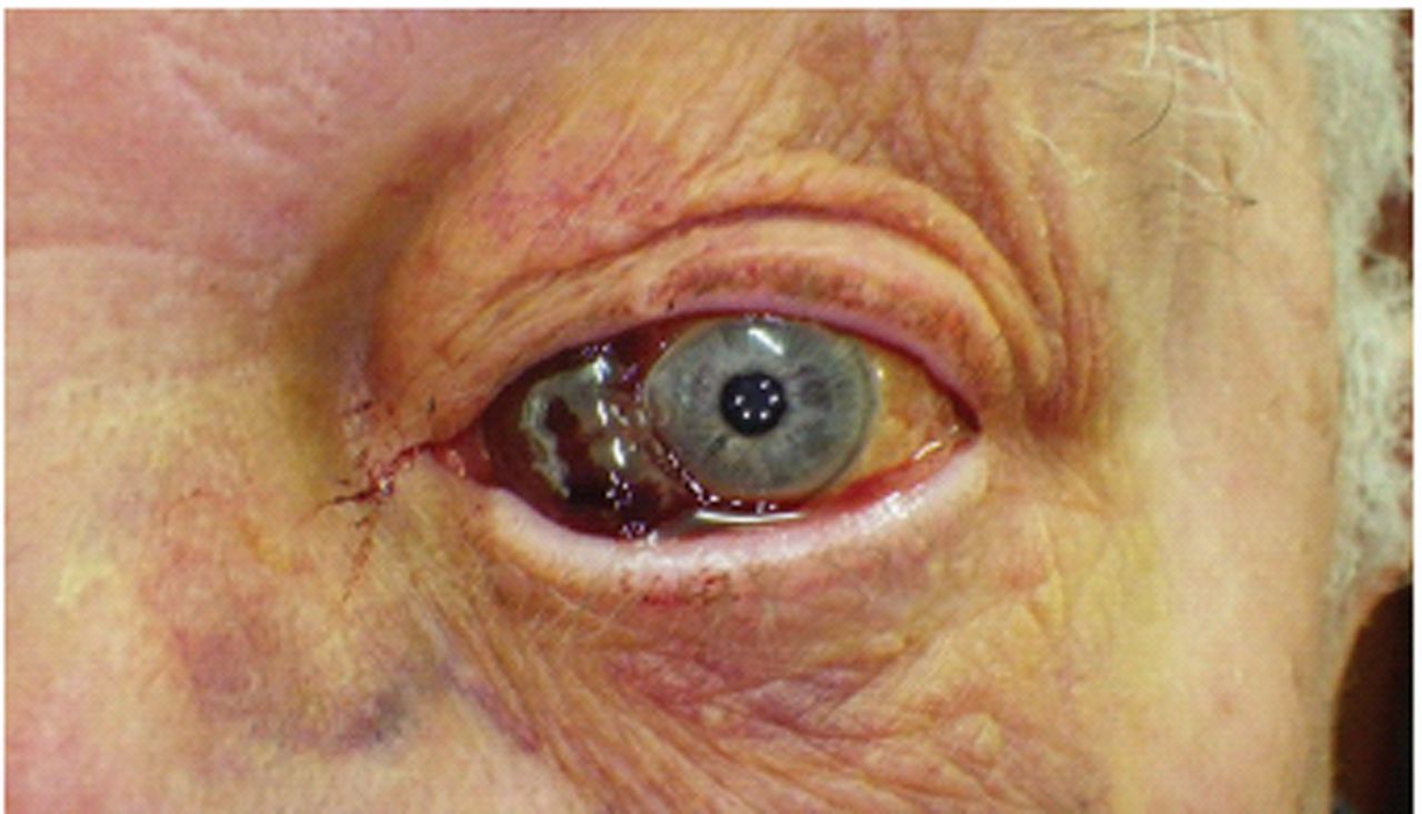
Severe subconjunctival haemorrhage in an older gentleman on warfarin and clopidogrel. Warfarin levels were checked and found to be normal. Thus reassurance only required. Note the five LED ring flash lights in the pupil.
At the very least, a head and shoulders photograph of every patient on the practice list is useful to confirm identity and to reduce the chance of errors among people having similar names. Images transmitted to GP surgeries by the police can help to prevent people obtaining fraudulent prescriptions, and the identities of ‘temporary resident’ patients can be confirmed by transmitted images. Photographs of patient’s injuries in domestic abuse cases are recommended rather than using simple verbal descriptions. Patients accept the need of having their photographs included on driving licences, bus passes, and security badges. It is unlikely that they would resist the introduction of photographs in their medical records.
Dermatology comprises a substantial part of the GP workload and this includes the evaluation of pigmented lesions. Those not demanding immediate referral can be photographed and the image used to check progress at follow-up appointments. Photographs are particularly useful too in the assessment of changes in gravitational ulcers. Sharing the images with patients may in these circumstances strengthen the understanding between clinician and patient. The value of photographs, before and after surgery has always been recognised in the field of plastic surgery. Their value across the specialties has been underexploited. Like dentists, doctors should appreciate the medico-legal advantages of photographic records.
Few doctors can resist the challenge of the photographic quiz that features in so many popular journals. A quick glance should be all that is necessary for the accurate diagnosis of a host of conditions, and this ability to make a ‘spot diagnosis’ a source of satisfaction among practitioners. We congratulate ourselves when we recognise the malar flush of mitral stenosis, the snarling facies of myasthenia gravis, and the gorilla-like face of acromegaly. But perhaps we were uncertain about the diagnosis of the patient showing the blue grey nose and cheeks, and sent off the image to someone we thought would know (the medical equivalent of phone a friend). That turned out to be a side effect of amiodarone. We could go on to show the image at a didactic practice meeting. From abducens nerve palsy to arthropathy, from valgus to vicar’s knee, the photograph offers immeasurable educational opportunities.
Like the routine measurement of height, weight, and blood pressure, the photographing of patients should not be a once and for all exercise. The comparison of photographs taken over time provides vivid evidence of the onset of disease and its progress. Photographs of people who live alone, and have no family to comment on their appearance, are particularly valuable. Perhaps even more precious are those of people who suffer from intellectual disability and whose verbal communication is restricted. Often they live in group homes where, because the care staff is constantly changing, no one is in any position to observe important changes in their appearance.
No one would deny the old maxim: a picture is worth a thousand words. We should then recognise this, and make use of our cameras, mobile phones, and iPads to capture for all time exactly what is before our eyes. We allow hardly any social event to escape our cameras. Why do we not capture the medical events in the lives of our patients? It’s so simple and so much fun too!

Phytophotodermatitis. A young man on a beach in Cornwall wrapped plant material around one of his feet as a substitute shoe. This fell off but plant juice on the dorsum of the foot under the action of sunlight caused a severe blistering eruption.
Notes
Provenance
Freely submitted; not externally peer reviewed.
Photographs and consent
Both photographs were taken with a simple-to-use compact, robust, Pentax Optio WG2 camera in digital microscope mode. Both photographs have been attached to the patient clinical record as the file size in this mode is only two megabytes, which is small. Both patients have provided consent for these images to be published.
- © British Journal of General Practice 2013
In this issue


Jump to section
More in this TOC Section
Related Articles
Cited By...

