


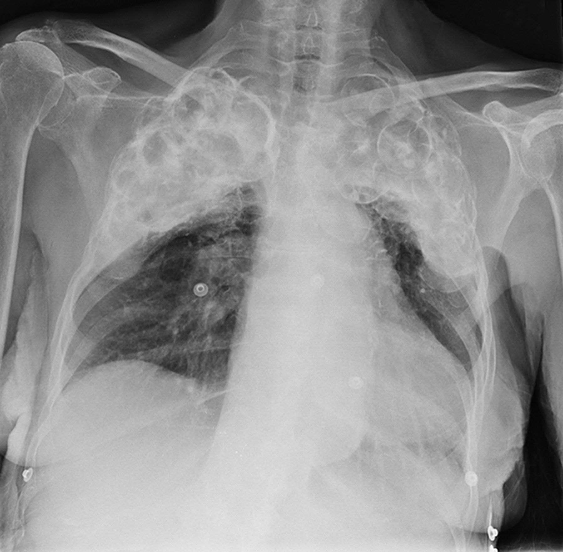
Imaging taken early last Autumn in the hospital I worked in revealed startling images. Multiple round ring shadows, overlapping against each other, were clearly visible in the apices of both lung fields in the chest radiograph of a patient who had just been newly admitted. The junior doctors involved in the immediate care of the patient requested an urgent report on the X-ray, which confirmed that the patient had been the recipient of ‘plombage’, a form of surgical therapy used for the treatment of tuberculosis (TB) before the 1950s and prior to the use of antituberculosis drugs; the opacities in this case being ping pong balls.
Plombage was a form of collapse therapy using foreign material aimed at treating localised lung disease1 primarily the parenchymal cavities, the main site at which bacterial proliferation occurs.2 It was thought that collapse would allow the lung to rest and aid healing by expunging the cavitating lesions.1 The first form of collapse therapy was performed by Hippocrates over 2400 years ago when he inserted a pig’s bladder into a patient’s chest and inflated it.3 Although the principle behind Hippocrates’ intervention was unknown, collapse therapy aimed specifically at the treatment of TB was first performed by Tuffier in 1891.3
Material used for plombage varied, from the use of omentum and lipoma by Tuffier, to silk, drawing crayons and lead bullets as reported by Archibald,3 Lucite balls (a form of thermoplastic acrylic resin) to the ping-pong balls in our patient.3 The therapy was popular as a one-stage procedure which demonstrated ‘excellent results’ in early trials.4 However, it soon became apparent this was not the complete picture when high complication rates were seen.
Horowitz et al 5 writes that the average time for complications to occur was 37 years after therapy and they were often related to migration of the exogenous material or to infection. In one case report, a 16-year-old noticed a ‘tender hard lump’ in the axilla 5 years after undergoing the procedure. Under the impression this was axillary adenitis, incision of the area eventually revealed a polythene ball which had migrated into the subcutaneous tissues.4 A second case of migration of plomb material described a patient who was re-admitted 18 months later with bowel obstruction. Laparotomy resulted in the removal of a Lucite sphere from the jejunum. Further investigation revealed a fistula which had allowed the ball to pass through the oesophagus into the lower gastrointestinal tract.5 Another case involved a patient who underwent plombage using paraffin wax masses and started coughing up large quantities of wax almost 28 years later.6

Today, with the advent of effective pharmacological therapy, surgical intervention for the treatment of tuberculosis is employed less commonly and if so, is mainly used for the resection of destroyed or infected lung.7,8 The patient admitted under our care had thankfully not suffered from any complications after her experience of this procedure and had remained free from TB. Her chest X-ray neatly demonstrates the novel ways in which our predecessors approached the management of this condition and how therapy for it has evolved since then.
Notes
Patient consent
The patient has consented to publication of this article and the associated image.
- © British Journal of General Practice 2014
In this issue


Jump to section
More in this TOC Section
Related Articles
Cited By...

