
Main menu

  • HOME
  • ONLINE FIRST
  • CURRENT ISSUE
  • ALL ISSUES
  • AUTHORS & REVIEWERS
  • SUBSCRIBE
  • RESOURCES
    • About BJGP
    • Conference
    • Advertising
    • BJGP Life
    • eLetters
    • Librarian information
    • Alerts
    • Resilience
    • Video
    • Audio
    • COVID-19 Clinical Solutions
  • RCGP
    • BJGP for RCGP members
    • BJGP Open
    • RCGP eLearning
    • InnovAIT Journal
    • Jobs and careers
    • RCGP e-Portfolio

User menu

  • Subscriptions
  • Alerts
  • Log in

Search

  • Advanced search
British Journal of General Practice
  • RCGP
    • BJGP for RCGP members
    • BJGP Open
    • RCGP eLearning
    • InnovAIT Journal
    • Jobs and careers
    • RCGP e-Portfolio
  • Subscriptions
  • Alerts
  • Log in
  • Follow bjgp on Twitter
  • Visit bjgp on Facebook
  • Blog
  • Listen to BJGP podcast
Advertisement
British Journal of General Practice

Advanced Search

  • HOME
  • ONLINE FIRST
  • CURRENT ISSUE
  • ALL ISSUES
  • AUTHORS & REVIEWERS
  • SUBSCRIBE
  • RESOURCES
    • About BJGP
    • Conference
    • Advertising
    • BJGP Life
    • eLetters
    • Librarian information
    • Alerts
    • Resilience
    • Video
    • Audio
    • COVID-19 Clinical Solutions
Clinical Intelligence

Peripheral arterial disease: diagnostic challenges and how photoplethysmography may help

Scott Wilkes, Gerard Stansby, Andrew Sims, Shona Haining and John Allen
British Journal of General Practice 2015; 65 (635): 323-324. DOI: https://doi.org/10.3399/bjgp15X685489
Scott Wilkes
Coquet Medical Group, Amble, Northumberland; professor of general practice and primary care, Department of Pharmacy, Health and Wellbeing, University of Sunderland, Sunderland.
Roles: GP Principal
  • Find this author on Google Scholar
  • Find this author on PubMed
  • Search for this author on this site
Gerard Stansby
Newcastle University and Newcastle Hospitals, Newcastle upon Tyne; chair of the North of England Clinical Network Vascular Surgery Group.
Roles: Professor of vascular surgery
  • Find this author on Google Scholar
  • Find this author on PubMed
  • Search for this author on this site
Andrew Sims
Clinical scientist and head of the Clinical Measurement and Engineering Unit, Medical Physics Department, Newcastle upon Tyne Hospitals NHS Foundation Trust; honorary senior lecturer, Institute of Cellular Medicine, Newcastle University, Newcastle upon Tyne.
  • Find this author on Google Scholar
  • Find this author on PubMed
  • Search for this author on this site
Shona Haining
North of England Commissioning Support, Newcastle.
Roles: Senior research and development manager
  • Find this author on Google Scholar
  • Find this author on PubMed
  • Search for this author on this site
John Allen
Microvascular Diagnostics Service in Medical Physics, Newcastle upon Tyne Hospitals NHS Foundation Trust; honorary clinical senior lecturer, Institute of Cellular Medicine, Faculty of Medical Sciences, Newcastle University, Newcastle upon Tyne.
Roles: Clinical scientist and clinical lead
  • Find this author on Google Scholar
  • Find this author on PubMed
  • Search for this author on this site
  • Article
  • Figures & Data
  • Info
  • eLetters
  • PDF
Loading

INTRODUCTION

Peripheral arterial disease (PAD) affects approximately 20% of patients aged ≥60 years.1 A GP with an average list size of 1800 patients can expect to have 50–60 patients with PAD. Ankle-brachial pressure index (ABPI), which is the ratio of the ankle to brachial systolic pressure measured by sphygmomanometer and hand-held Doppler ultrasound probe, is used to assess PAD in general practice. ABPI has been shown to have a sensitivity of 95% and specificity of 99% compared to angiographic imaging,2 however it is relatively time-consuming and requires adequately trained staff. There are limitations with ABPI in patients with diabetes, renal disease, and older people where an underestimation of disease can occur with a falsely high ratio due to the presence of incompressible calcified blood vessels.

Clinical Question

Can peripheral arterial disease be accurately diagnosed in general practice?

DIAGNOSTIC CHALLENGES

The characteristic clinical diagnostic feature of PAD is intermittent claudication (IC), muscle pain brought on by exercise, relieved by rest and experienced in the calf with femoropopliteal disease and in the buttock, hip or thigh with aortoiliac disease. However it is sometimes difficult to distinguish clinically between claudication and leg pain from other causes such as spinal stenosis.

ABPI methodology

The first problem we face is that a significant proportion of patients in general practice who are arteriopaths are asymptomatic and ABPI has been routinely adopted to confirm and quantify the diagnosis of peripheral vascular disease in those with IC. Unfortunately the exact methodology is not standardised, beginning with a choice of three arteries in the ankle each with potentially differing pressures and some of which are commonly congenitally absent. Similarly the clinician has a choice of two arms for the brachial pressure and there is no consensus on how the central reference pressure should be assessed. Indeed, arteriosclerosis is a systemic disease. Failure to lie supine can introduce a hydrostatic pressure difference between arm and leg. A number of environmental factors such as ambient temperature can also affect peripheral vasomotor tone. The occlusive cuff method itself has been called into question with a likely underestimate of true pressure drop varying with site of lesion and which may be exacerbated with baseline hypotension or hypertension confounding ABPI readings. Finally, there is no consensus on normal ranges for ABPI ratios, and hence referral thresholds, evidenced by its absence in the most recent National Institute for Health and Care Excellence guidance.3

Limitations of ABPI

As well as methodological limitations and despite the claimed 95% sensitivity and 99% specificity, there are a number of problematic assumptions. Many of the studies of ABPI have used control subjects that are clinically asymptomatic volunteers rather than angiographically proven normals. A rich collateral vessel network may be responsible for a proportion of asymptomatic PAD patients and ABPI would fail to unmask this cohort. Heavy arterial calcification resulting in incompressible arteries in diabetic, renal, and older patients renders the ABPI non-diagnostic. This is also problematic in rarer conditions such as systemic sclerosis and rheumatic diseases. There is a need for a non-invasive alternative to ABPI for assessing peripheral vascular disease in general practice.

PHOTOPLETHYSMOGRAPHY

Photoplethysmography (PPG) is offering a new look at diagnosis and management of PAD in general practice. PPG is a low cost, non-invasive, simple-to-use, and fast- to-perform optical technique based on peripheral pulse waveform analysis and has been shown to have good sensitivity and specificity for PAD when compared to ABPI.4 However the place of this technology in general practice is yet to be established.

Methodology and challenges

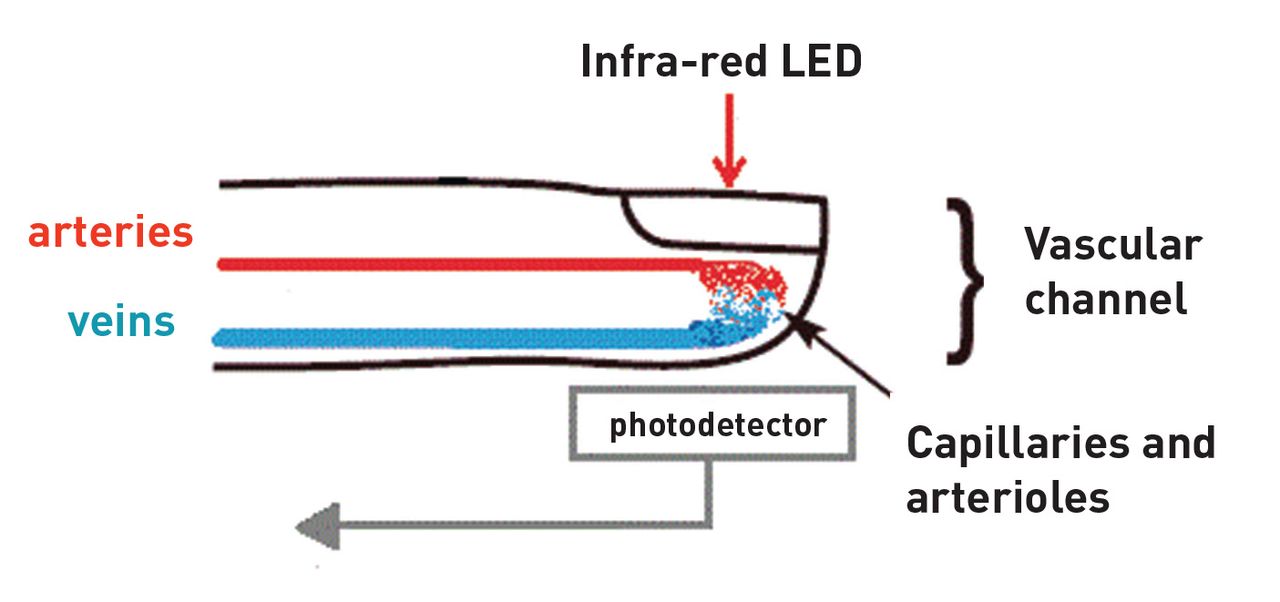
PPG typically uses near-infrared (or red) light to measure the changes in tissue blood volume associated with each heartbeat.5 In its simplest form a light source is directed towards the tissue microvascular bed, for example to the ear lobe, finger, or toe pad, and an adjacent photodetector measures the small changes in light absorption to form a PPG pulse waveform (Figure 1). Although the exact nature of PPG is not fully understood its signatures in health and vascular disease have been described and reviewed by Allen.5

Figure 1.
  • Download figure
  • Open in new tab
  • Download powerpoint
Figure 1.

Photoplethysmography.

The waveforms from PPG technology are typically pulsatile and can be compared between sites such as the toes and fingers. In PAD a damping effect is produced with a reduction in amplitude and changes in waveform shape and timing compared to a healthy vascular bed.

A simpler variant of PPG, pulse oximetry, has already been successfully deployed in general practice. The next stage in the development of this technology is in its routine use for PAD patients in general practice.

WHAT IT MEANS FOR GENERAL PRACTICE

The key determinant to the successful embedding of advanced PPG technology in general practice is the ability to produce clear results indicating significant or non-significant disease and for it to be reliably delivered by a range of primary care professionals. The purpose of such technology will be:

  • to identify a cohort of patients in whom there is no significant PAD and who can be reassured;

  • to identify a cohort of patients in whom there is significant PAD and make onward referral for further evaluation and/or intervention; and

  • to identify a cohort of patients with mild PAD where primary care management is indicated (Box 1).

Box 1.

Summary of PAD management and annual monitoring in general practice

  • Assessment of symptoms

  • Ankle-brachial pressure index (ABPI) estimation

  • Smoking cessation advice

  • Cholesterol lowering

  • Blood pressure management

  • Weight management and exercise

  • HBA1c estimation

  • Anti-platelet treatment

PAD is a window on widespread atherosclerotic cardiovascular disease6 and advanced PPG technology may give opportunities for cardiovascular screening in the future.

FURTHER WORK

With support from the National Institute for Health Research Invention for Innovation scheme, the Novel Test Evaluating Peripheral Arterial Disease (NOTEPAD) project will develop a PPG device for use in primary care. It remains to be seen whether PPG technology can produce an output that guides the management of PAD in general practice. Technological advances in PAD management have lagged behind in comparison with coronary heart disease and stroke management. It is the intention of the NOTEPAD investigators to develop and demonstrate the worth of this technology to the primary care clinician.

Notes

Funding

This report is independent research funded by the National Institute for Health Research (Invention for Innovation, ‘Innovative photoplethysmography technology for rapid noninvasive assessment of peripheral arterial disease in primary care,’ II-C1-0412-20003). Views expressed in this publication are those of the authors and not necessarily those of the NHS, the NIHR or the Department of Health.

Provenance

Freely submitted; not externally peer reviewed.

Competing interests

John Allen and Andrew Simms are employed by The Newcastle upon Tyne Hospitals NHS Foundation Trust which holds the majority rights to the intellectual property of the NOTEPAD technology.

Discuss this article

Contribute and read comments about this article: bjgp.org/letters

  • Received February 24, 2015.
  • Accepted February 25, 2015.
  • © British Journal of General Practice 2015

REFERENCES

  1. 1.↵
    1. Fowkes FG,
    2. Housley E,
    3. Cawood EH,
    4. et al.
    (1991) Edinburgh Artery Study: prevalence of asymptomatic and symptomatic peripheral arterial disease in the general population. Int J Epidemiol 20(2):384–392.
    OpenUrlCrossRefPubMed
  2. 2.↵
    1. Bernstein EF,
    2. Fronek A
    (1982) Current status of noninvasive tests in the diagnosis of peripheral arterial disease. Surg Clin North Am 62(3):473–487.
    OpenUrlPubMed
  3. 3.↵
    1. National Institute for Health and Care Excellence
    Lower limb peripheral arterial disease: diagnosis and management, http://www.nice.org.uk/guidance/cg147 (accessed 28 Apr 2015), p CG147.
  4. 4.↵
    1. Allen J,
    2. Overbeck K,
    3. Nath AF,
    4. et al.
    (2008) A prospective comparison of bilateral photoplethysmography versus the ankle-brachial pressure index for detecting and quantifying lower limb peripheral arterial disease. J Vasc Surg 47(4):794–802.
    OpenUrlCrossRefPubMed
  5. 5.↵
    1. Allen J
    (2007) Photoplethysmography and its application in clinical physiological measurement. Physiol Meas 28(3):R1–39.
    OpenUrlCrossRefPubMed
  6. 6.↵
    1. Doobay AV,
    2. Anand SS
    (2005) Sensitivity and specificity of the ankle-brachial index to predict future cardiovascular outcomes: a systematic review. Arterioscler Thromb Vasc Biol 25(7):1463–1469.
    OpenUrlAbstract/FREE Full Text
Back to top
Previous ArticleNext Article

In this issue

British Journal of General Practice: 65 (635)
British Journal of General Practice
Vol. 65, Issue 635
June 2015
  • Table of Contents
  • Index by author
Download PDF
Download PowerPoint
Article Alerts
Or,
sign in or create an account with your email address
Email Article

Thank you for recommending British Journal of General Practice.

NOTE: We only request your email address so that the person to whom you are recommending the page knows that you wanted them to see it, and that it is not junk mail. We do not capture any email address.

Enter multiple addresses on separate lines or separate them with commas.
Peripheral arterial disease: diagnostic challenges and how photoplethysmography may help
(Your Name) has forwarded a page to you from British Journal of General Practice
(Your Name) thought you would like to see this page from British Journal of General Practice.
CAPTCHA
This question is for testing whether or not you are a human visitor and to prevent automated spam submissions.
Citation Tools
Peripheral arterial disease: diagnostic challenges and how photoplethysmography may help
Scott Wilkes, Gerard Stansby, Andrew Sims, Shona Haining, John Allen
British Journal of General Practice 2015; 65 (635): 323-324. DOI: 10.3399/bjgp15X685489

Citation Manager Formats

  • BibTeX
  • Bookends
  • EasyBib
  • EndNote (tagged)
  • EndNote 8 (xml)
  • Medlars
  • Mendeley
  • Papers
  • RefWorks Tagged
  • Ref Manager
  • RIS
  • Zotero

Share
Peripheral arterial disease: diagnostic challenges and how photoplethysmography may help
Scott Wilkes, Gerard Stansby, Andrew Sims, Shona Haining, John Allen
British Journal of General Practice 2015; 65 (635): 323-324. DOI: 10.3399/bjgp15X685489
del.icio.us logo Digg logo Reddit logo Twitter logo CiteULike logo Facebook logo Google logo Mendeley logo
  • Tweet Widget
  • Facebook Like
  • Google Plus One
  • Mendeley logo Mendeley

Jump to section

  • Top
  • Article
    • INTRODUCTION
    • DIAGNOSTIC CHALLENGES
    • PHOTOPLETHYSMOGRAPHY
    • WHAT IT MEANS FOR GENERAL PRACTICE
    • FURTHER WORK
    • Notes
    • REFERENCES
  • Figures & Data
  • Info
  • eLetters
  • PDF

More in this TOC Section

  • How to manage low testosterone level in men: a guide for primary care
  • Hyperparathyroidism (primary) NICE guideline: diagnosis, assessment, and initial management
  • The atypical presentation of COVID-19 as gastrointestinal disease: key points for primary care
Show more Clinical Intelligence

Related Articles

Cited By...

Advertisement

BJGP Life

BJGP Open


@BJGPjournal's Likes on Twitter


British Journal of General Practice

NAVIGATE

  • Home
  • Current Issue
  • All Issues
  • Online First
  • Authors & reviewers

RCGP

  • BJGP for RCGP members
  • BJGP Open
  • RCGP eLearning
  • InnovAiT Journal
  • Jobs and careers
  • RCGP e-Portfolio

MY ACCOUNT

  • RCGP members' login
  • Subscriber login
  • Activate subscription
  • Terms and conditions

NEWS AND UPDATES

  • About BJGP
  • Alerts
  • RSS feeds
  • Facebook
  • Twitter

AUTHORS & REVIEWERS

  • Submit an article
  • Writing for BJGP: research
  • Writing for BJGP: other sections
  • BJGP editorial process & policies
  • BJGP ethical guidelines
  • Peer review for BJGP

CUSTOMER SERVICES

  • Advertising
  • Contact subscription agent
  • Copyright
  • Librarian information

CONTRIBUTE

  • BJGP Life
  • eLetters
  • Feedback

CONTACT US

BJGP Journal Office
RCGP
30 Euston Square
London NW1 2FB
Tel: +44 (0)20 3188 7679
Email: journal@rcgp.org.uk

British Journal of General Practice is an editorially-independent publication of the Royal College of General Practitioners
© 2021 British Journal of General Practice

Print ISSN: 0960-1643
Online ISSN: 1478-5242