


INTRODUCTION
Erythema multiforme (EM) is an acute and self-limiting hypersensitivity reaction. This article describes the case of a patient who suffered from a chest infection and later presented with the mucosal manifestations of EM. This condition manifests as skin lesions, most commonly in reaction to herpes simplex virus (HSV) 1 and 2, Mycoplasma pneumoniae, or certain medications.1
CASE REPORT
A 31-year-old male patient presented to his GP with cough and fever, sores in his mouth, and itchy eyes. He had not received any medical attention prior to this appointment, nor had he taken any recent medications. He was initially prescribed aciclovir to treat oral herpes simplex infection, amoxicillin for chest infection, and chloramphenicol for conjunctivitis. Two weeks later he was seen by the medical assessment unit. In addition to blisters on his mouth and lips (Figure 1), he developed pruritic blisters on his penis and foreskin (Figure 2), small lesions acrally, and red eyes. On this basis the consultant medical physician referred the patient to the genitourinary medicine (GUM) clinic to rule out sexually-acquired reactive arthritis (SARA) secondary to chlamydia infection.
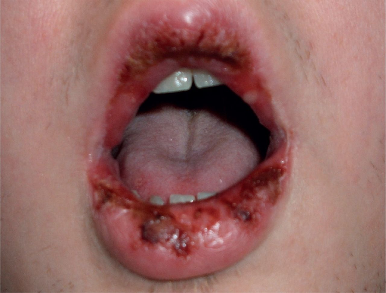
Erosions of the oral mucosa in a case of erythema multiforme.

Erythema multiforme-associated mucosal erosions of the glans penis.
The GUM consultant made a provisional diagnosis of Stevens–Johnson syndrome (SJS) and referred the patient to a dermatologist.
When reviewed by the dermatologist, the patient’s symptoms had improved and he was able to swallow and drink as normal. He had no recollection of having cold sores or medications prior to the onset of his symptoms.
With this in mind, EM with mucosal involvement secondary to M. pneumoniae infection was considered and this was later confirmed via the serological particle agglutination test (>1:640, indicating ongoing infection). No specific treatment was prescribed, although Vaseline® for his lips was advised to stop them from sticking.
DISCUSSION
This case demonstrates an interesting scenario in which the initial diagnosis was unclear to both GP and secondary care physician, and a range of reasonable differential diagnoses were made. Mouth ulceration secondary to herpes simplex and conjunctivitis are both common localised infections.2,3 SARA presents as a triad of urethritis, arthritis, and conjunctivitis, or a pentad including circinate balanitis and keratoderma blenorrhagicum.4 Circinate balanitis in this case is an appropriate differential with similar appearances to the genital mucosal erosions in EM.
The most common aetiological agents of EM are HSV 1 and 2, which account for >50% of cases. M. pneumoniae is the second most common infective agent (particularly in children), followed by fungal infections and other non-herpes viral infections. The most associated medications with EM are barbiturates, Non-steroidal anti-inflammatory drugs (NSAIDs), penicillins, phenothiazines, sulfonamides, and hydantoins. Radiotherapy is another known trigger.1
Disease re-classification in 1993 clarified the distinguishing clinical features between EM, SJS, and toxic epidermal necrolysis (TEN). Although they resemble a disease spectrum, these diagnoses vary in the pattern of skin lesions and degree of epidermal detachment. These terms are now used in preference over the old terminology: EM minor and EM major.5
PRESENTATION, DIAGNOSIS, AND MANAGEMENT
EM is a hypersensitivity reaction. Patients present with characteristic ‘target’ rashes with or without mucosal involvement. They may describe prodromal symptoms and often complain of itching or burning at the sites of the lesions.6
Initial EM lesions appear as polymorphous macules that develop into papules and sometimes large plaques. Concentric colour zones are characteristic of ‘target’ lesions, which vary in morphology (hence the name erythema multiforme). EM is clinically diagnosed by the presence of these typical target lesions, as well as raised atypical targets, which most often erupt acrally in EM (as was the case in this patient). Skin biopsies are not necessary unless the diagnosis is unclear. Mucosal erosions may also be present in oral, genital, or ocular regions. Epidermal detachment is confined to <10% of the body surface.
In contrast to EM, SJS has no typical target lesions, flat atypical targets, and confluent purpuric macules on the face and trunk with severe mucosal erosions at one or more mucosal sites. As with EM, epidermal detachment is limited to <10% of the body surface.
TEN is similar to SJS in that there are no typical targets but flat atypical targets. However, the disease appears with severe mucosal erosion and progresses to diffuse generalised detachment of the epidermis to >30% of the body surface area.6
Management is directed towards treating symptoms and removing the cause, and therefore to treat the suspected infection or to discontinue the causative medication.5 Furthermore, identifying the aetiology can provide valuable information regarding the prognosis. If the trigger is due to mycoplasma or non-herpes simplex viral infection, then it is usually a one-off and the episode will resolve spontaneously within 3–5 weeks without sequelae. However, HSV is known to cause recurrent EM, which can be treated with continuous oral aciclovir (400 mg twice daily).5,7,8 When suppressive antivirals are not effective, patients should be referred to dermatology.1
CONCLUSION
This case highlights the need to keep an open mind that dermatological conditions can be due to local or systemic processes. It is important to take a full and thorough history to identify potential mediators of EM, and to fully examine the skin to rule out rashes elsewhere. Although 40% of causes are idiopathic, there are clear links with certain infections and medications (as is the case in this scenario), and these should be identified for treatment and prognostic purposes. Not all genital ulcers are related to genital infections and therefore it is important to bear in mind systematic and dermatological diseases that may appear in the genital region.
Notes
Patient consent
The patient gave consent for publication of this case report and images.
Provenance
Freely submitted; not externally peer reviewed.
Competing interests
The authors have declared no competing interests.
Discuss this article
Contribute and read comments about this article: bjgp.org/letters
- Received September 9, 2015.
- Revision requested September 24, 2015.
- Accepted September 29, 2015.
- © British Journal of General Practice 2016
In this issue


Jump to section
More in this TOC Section
Related Articles
Cited By...

