Article Text

Abstract
Objective: To determine the performance of a new NT-proBNP assay in comparison with brain natriuretic peptide (BNP) in identifying left ventricular systolic dysfunction (LVSD) in randomly selected community populations.
Methods: Blood samples were taken prospectively in the community from 591 randomly sampled individuals over the age of 45 years, stratified for age and socioeconomic status and divided into four cohorts (general population; clinically diagnosed heart failure; patients on diuretics; and patients deemed at high risk of heart failure). Definite heart failure (left ventricular ejection fraction (LVEF) < 40%) was identified in 33 people. Samples were handled as though in routine clinical practice. The laboratories undertaking the assays were blinded.
Results: Using NT-proBNP to diagnose LVEF < 40% in the general population, a level of > 40 pmol/l had 80% sensitivity, 73% specificity, 5% positive predictive value (PPV), 100% negative predictive value (NPV), and an area under the receiver-operator characteristic curve (AUC) of 76% (95% confidence interval (CI) 46% to 100%). For BNP to diagnose LVSD, a cut off level of > 33 pmol/l had 80% sensitivity, 88% specificity, 10% PPV, 100% NPV, and AUC of 88% (95% CI 75% to 100%). Similar NPVs were found for patients randomly screened from the three other populations.
Conclusions: Both NT-proBNP and BNP have value in diagnosing LVSD in a community setting, with similar sensitivities and specificities. Using a high cut off for positivity will confirm the diagnosis of LVSD but will miss cases. At lower cut off values, positive results will require cardiac imaging to confirm LVSD.
- AUC, area under receiver-operator characteristic curve
- BNP, brain natriuretic peptide
- ECHOES, echocardiographic Heart of England screening study
- LR, likelihood ratio
- LVEF, left ventricular ejection fraction
- LVSD, left ventricular systolic dysfunction
- NPV, negative predictive value
- PPV, positive predictive value
- natriuretic peptides
- left ventricular systolic dysfunction
- heart failure
Statistics from Altmetric.com
- AUC, area under receiver-operator characteristic curve
- BNP, brain natriuretic peptide
- ECHOES, echocardiographic Heart of England screening study
- LR, likelihood ratio
- LVEF, left ventricular ejection fraction
- LVSD, left ventricular systolic dysfunction
- NPV, negative predictive value
- PPV, positive predictive value
Left ventricular systolic dysfunction (LVSD) is an increasingly common disorder,1 with a prevalence of around 2%.2,3 It has a poor prognosis4 and is associated with a reduced quality of life,5 especially where it is the cause of heart failure. As outcomes in heart failure and LVSD are linked to the stage of disease, early and accurate diagnosis is needed to guide appropriate management.
Even in symptomatic patients, the diagnosis of LVSD is not possible on clinical grounds alone, as objective evidence of cardiac dysfunction is required6,7; this is usually obtained from echocardiographic studies. A reliable diagnosis of LVSD is important because several treatments, especially the use of angiotensin converting enzyme (ACE) inhibitors8 and β blockers,9 improve symptoms, quality of life, and disease prognosis, and reduce health care use and costs. However, most patients with symptomatic LVSD are presumed to have LVSD on clinical grounds alone, without further investigation.10 Furthermore, early diagnosis may also be important in asymptomatic LVSD, as treatment can delay or reverse disease progression and the onset of heart failure.11,12 Diagnostic tools may therefore need to encompass screening strategies13,14 as well as case identification in symptomatic patients.
LVSD is usually investigated by echocardiography, which provides a semi-objective assessment of ventricular and valve function. However, access to echocardiography is limited in many health care systems,11 especially in primary care, and therefore the validity of a clinical diagnosis of LVSD in primary care is often poor when patients are assessed against objective criteria.15 The ECG has been advocated as an easy non-invasive method for diagnosing LVSD, as a normal recording will, in most cases, exclude left ventricular dysfunction.16,17 However, many non-specialists18 are unreliable at assessing ECG changes.
Potential new diagnostic aids are natriuretic peptide assays. Brain natriuretic peptide (BNP) is a 32 amino acid biologically active peptide, cleaved from the 108 amino acid proBNP, released from the cardiac ventricles in response to chamber stretch. BNP has vasodilator and natriuretic properties. The second remnant following cleavage, N-terminal proBNP (NT-proBNP), is a 76 amino acid with no known biological function.
BNP showed a high sensitivity and modest specificity in confirming LVSD in 1653 adults (aged 25–75 years) screened in an area of high cardiovascular disease19 and in 155 elderly patients over 75 years screened in primary care.20 NT-proBNP has been less investigated, but the assay used in this study reported a sensitivity of 100%, a specificity of 70%, a positive predictive value (PPV) of 7%, a negative predictive value (NPV) of 100%, and an area under the receiver-operator characteristic (ROC) curve of 92% (95% confidence interval (CI) 82% to 100%) in the diagnosis of heart failure in the ECHOES (echocardiographic Heart of England screening) cohort.21 In this paper we report complementary original data on the value of the NT-proBNP assay in the diagnosis of LVSD in various community populations and compare the data with the performance of the most commonly used BNP assay.
METHODS
This was a prospective substudy of the ECHOES study of LVSD and heart failure prevalence.2 The main study was carried out in 16 randomly selected primary care practice populations in England following stratification for age and socioeconomic status. Patients were randomly selected from each of four discrete population cohorts, identified in each practice from computerised practice registers, comprising:
-
randomly sampled patients from those aged 45 years and older (general population screen)
-
patients with a clinical (unvalidated) diagnostic label of heart failure
-
patients prescribed diuretic drugs
-
patients at high risk of heart failure (history of previous myocardial infarction, angina, hypertension, or diabetes).
This natriuretic peptide substudy was conducted in the last four practices, containing the final 607 consecutively screened subjects randomly selected from the four target groups (307 in the general population, 103 with an existing clinical diagnosis of heart failure, 87 taking diuretics, and 134 at high risk of heart failure, with some sampled from more than one cohort). These practices were representative of the socioeconomic spread of patients achieved by the initial stratification.
All patients were screened by history, New York Heart Association (NYHA) functional class, clinical examination, quality of life (short form 36 item (SF-36) health status questionnaire), spirometry, resting 12 lead ECG, and echocardiography, including Doppler studies. LVSD was defined as an ejection fraction under 40% by an area–length method from the apical four chamber view.22
Blood samples for natriuretic peptide assay were taken from the right antecubital fossa of consenting subjects after five minutes of supine rest into 5 ml K+EDTA tubes. Blood was kept at room temperature for up to 24 hours before transport to a local laboratory for centrifugation and freezing of plasma to −20°C. Once a week, the frozen samples were collected for central storage at −70°C. Assays of plasma BNP (Shinogi, Japan) and NT-proBNP (Roche Diagnostics, Germany) were subsequently done at a central laboratory, blinded to the screening assessments. The BNP immunoradiometric assay (IRMA) requires several operator dependent steps, including sample extraction, with total incubation of 18 to 22 hours. The NT-proBNP immunoassay (enzyme linked immunosorbent assay (ELISA)) requires three operator dependent steps and total incubation of under two hours.
Analysis
The diagnostic performance of BNP was assessed using ROC curves,23 formed by plotting sensitivity on the y axis and 1−specificity on the x axis for all possible cut off values of each diagnostic test. The overall discriminatory ability of the tests is demonstrated by the area under the curve (AUC).24 In addition to the AUCs, we identified the cut off value from the general population which maximised sensitivity without much loss of specificity. Likelihood ratios were also calculated, as a measure of the extent to which the pretest odds are altered by the test results (values > 1 increase the odds, values < 1 decrease the odds). The cut off values were then applied to the groups with a previous history of heart failure, diuretic use, and at high risk of heart failure. The resulting positive predictive values (PPV), negative predictive values (NPV), likelihood ratios (LR), and confidence intervals (CI) are presented.
The Hanley and McNeil method24 was used to compare the ROC curves of the two assays. Statistical analyses were carried out using SPSS and Microsoft EXCEL.
RESULTS
Of 607 eligible subjects, 591 (97%) consented to supply a blood sample. The basic demographics of the population screened are listed in table 1 and overall performance characteristics of the two assays and of the ECG in table 2.
Characteristics of total and left ventricular dysfunction (LVSD) populations
Area under receiver operator characteristic (ROC) curve, sensitivity, specificity, positive and negative predictive values, and likelihood ratios for brain natriuretic peptide and NT-proBNP in the diagnosis of left ventricular systolic dysfunction
Definite left ventricular systolic dysfunction, defined as a validated ejection fraction of less than 40%, was detected in 33 people from the four diagnostic groups: five from the general population, 21 from those with an existing heart failure diagnosis, seven from those on diuretic treatment, and 10 in the high risk group (10 patients were in more than one group).
Diagnosis of left ventricular systolic dysfunction using NT-proBNP
Using NT-proBNP to diagnose LVSD (LVEF < 40%), in the general population (fig 1), a cut off level of > 40 pmol/l had a sensitivity of 80% (95% CI 28% to 100%), a specificity of 73% (95% CI 68% to 78%), a PPV of 5% (95% CI 1% to 12%), an NPV of 100% (95% CI 98% to 100%), an LR+ of 2.9 (95% CI1.8 to 4.7), an LR− of 0.3 (95% CI 0.05 to 1.6), and an AUC of 0.76 (95% CI 0.46–1.0).
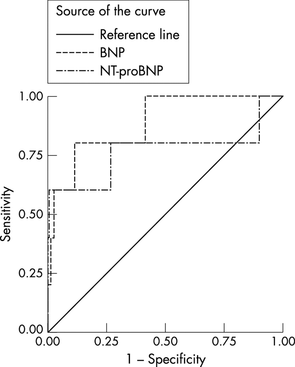
Receiver-operator characteristic (ROC) curves for brain natriuretic peptide (BNP) and N-terminal proBNP (NT-proBNP) in the diagnosis of definite left ventricular systolic dysfunction (left ventricular ejection fraction < 40%) in the general population.
Diagnosis of left ventricular systolic dysfunction using brain natriuretic peptides
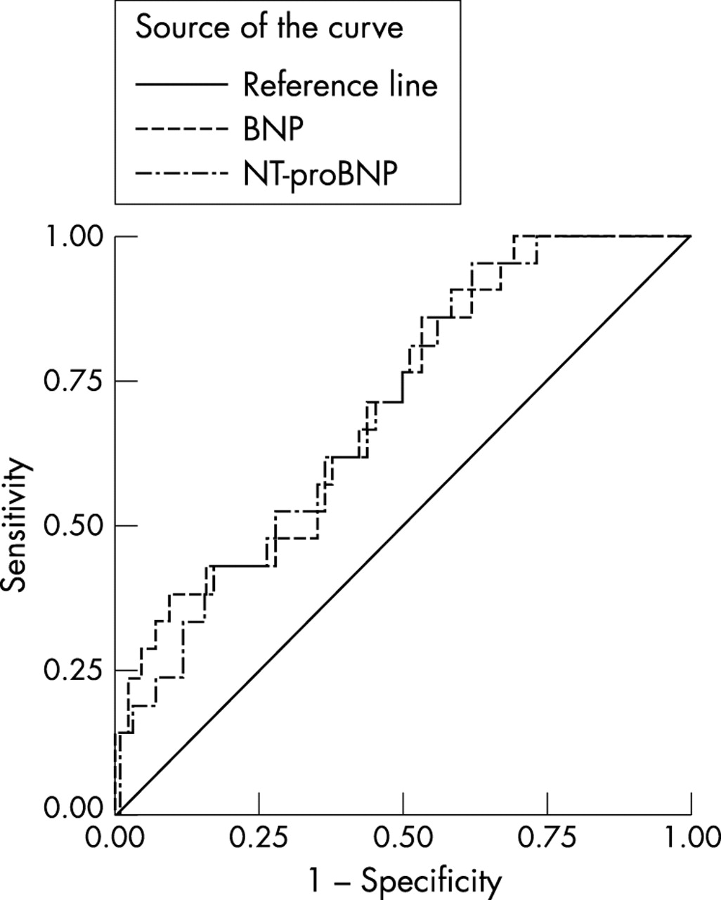
For BNP, a cut off of > 33 pmol/l had a sensitivity of 80% (95% CI 28% to 100%), a specificity of 88% (95% CI 84% to 92%), a PPV of 10% (95% CI 3% to 24%), an NPV of 100% (95% CI 98% to 100%), an LR+ of 6.7 (95% CI 3.9 to 11.5), an LR− of 0.2 (95% CI 0.04 to 1.3), and an AUC of 0.88 (95% CI 0.75 to 1.0) (fig 2). Similar performance characteristics were found using the same cut off values for the other population cohorts (figs 3 and 4), the NPVs, ranging from 88% to 100% and LR− from 0 to 0.75. The differences between the AUCs of the two natriuretic peptides were not significant in the diagnosis of LVSD.

Receiver-operator characteristic (ROC) curves for brain natriuretic peptide (BNP) and N-terminal proBNP (NT-proBNP) in the diagnosis of definite left ventricular systolic dysfunction (left ventricular failure) in the population with an established clinical diagnosis of heart failure.

Receiver-operator characteristic (ROC) curves for brain natriuretic peptide (BNP) and N-terminal proBNP (NT-proBNP) in the diagnosis of definite left ventricular systolic dysfunction (left ventricular failure) in the population on diuretics.

Receiver-operator characteristic (ROC) curves for brain natriuretic peptide (BNP) and N-terminal proBNP (NT-proBNP) in the diagnosis of definite left ventricular systolic dysfunction (left ventricular failure) in the high risk population.
DISCUSSION
In contrast to earlier studies of BNP assay performance in select patient groups, we believe these are the first data to compare the performance characteristics of both BNP and NT-proBNP assays in the diagnosis of LVSD, to compare the performance of the assays with that of ECGs, and to examine the assays in two populations: a large generalisable series of randomly selected adults with a validated LVSD diagnosis; and a comparator normative population, randomly selected from the same populations as the cases. The assays were also used under the same conditions and constraints as pertain to routine clinical practice. This “real world” study also sought to minimise the main confounding variables of diagnostic studies—namely, spectrum bias (the performance characteristics are presented separately for the different risk populations), verification bias, treatment paradox (any treatment effect will have diluted the size of the test performance), and reference standard error.
Both BNP and NT-proBNP assays were shown to be of value in diagnosing LVSD within a representative community based general adult population and also within at risk populations, with AUCs of at least 0.7.
The natriuretic assays were handled as they would be in everyday practice of primary care, namely collection by nurses and storage at room temperature for hours, so they should be more generalisable than earlier studies with specialised in-hospital handling of samples, including immediate chilled centrifugation and rapid freezing of samples to −70°C. Furthermore, the assays appear to be useful despite the influence of treatment on the peptide values,25 which could reduce the value of the assays in all but newly presenting patients.26
These data extend the positive conclusions of our earlier study reporting the performance characteristics of the NT-proBNP and BNP assays in the diagnosis of heart failure. With the high negative predictive values of both the BNP assays reported in this study, LVSD can effectively be ruled out at assay concentrations below our suggested cut off values. Such test characteristics suggest that the most appropriate use of the assays in routine practice would be as exclusion tests, where positive values would result in further cardiac investigation, including echocardiography. Choosing a higher cut off would become diagnostic of LVSD, but at the expense of missing cases.
The use of the assays as a triage test leading to echocardiography, as proposed above, would be useful in clinical practice because cardiac imaging would stage the degree of LVSD as well as confirming the diagnosis, thus guiding treatment intensity. Such a “pre-screening” use of the tests is analogous to cervical cytology in determining referral for colposcopy in cervical cancer screening. However, this interpretation of our data on the use of natriuretic peptide assays in LVSD diagnosis should be formally tested by cost–effectiveness trials of the assays in symptomatic and high risk screening strategies.
Acknowledgments
The ECHOES study was funded by the NHS R&D cardiovascular disease and stroke programme (FDRH and MKD were joint principal investigators). The costs of collecting and handling the assays were supported by an NHS Regional R&D grant, and the assays were donated by Roche Diagnostic Solutions.
FDRH and MKD were joint principal investigators and grant holders on the main study, with FDRH grant holder of this substudy. FDRH led the design and execution of the study and analysis. RCD conducted the majority of the clinical assessments and reporting, and made a major contribution to analysis. JEK and RH contributed to study design and analysis and overall study management. AKR performed statistical analysis. All authors contributed to drafts of the paper.
Competing interests: FDRH is a member of the European Society of Cardiology (ESC) Working Party on Heart Failure, is Chair of the British Primary Care Cardiovascular Society (PCCS), and Treasurer of the British Society for Heart Failure. MKD is Chairman of the British Society for Heart Failure. FDRH and MKD have received travel sponsorship and honoraria from a number of biotechnology and pharmaceutical companies with cardiovascular products for plenary talks and attendance at major cardiology scientific congresses and conferences.