Article Text

Statistics from Altmetric.com
Synopsis of recommendations
A summary of the initial management of patients admitted to hospital with suspected community acquired pneumonia (CAP) is presented in fig 8. Tables 4 and 5, respectively, summarise (1) the relevant microbiological investigations and (2) empirical antibiotic choices recommended in patients with CAP.

Hospital management of community acquired pneumonia (CAP) in the first 4 h. CXR, chest x ray; DBP, diastolic blood pressure; SBP, systolic blood pressure.
Recommendations for the microbiological investigation of community acquired pneumonia (CAP)
Initial empirical treatment regimens for community acquired pneumonia (CAP) in adults
Investigations (Section 5)
When should a chest radiograph be performed in the community?
It is not necessary to perform a chest radiograph in patients with suspected CAP unless:
The diagnosis is in doubt and a chest radiograph will help in a differential diagnosis and management of the acute illness. [D]
Progress following treatment for suspected CAP is not satisfactory at review. [D]
The patient is considered at risk of underlying lung pathology such as lung cancer. [D]
When should a chest radiograph be performed in hospital?
All patients admitted to hospital with suspected CAP should have a chest radiograph performed as soon as possible to confirm or refute the diagnosis. [D] The objective of any service should be for the chest radiograph to be performed in time for antibiotics to be administered within 4 h of presentation to hospital should the diagnosis of CAP be confirmed.
When should the chest radiograph be repeated during recovery?
The chest radiograph need not be repeated prior to hospital discharge in those who have made a satisfactory clinical recovery from CAP. [D]
A chest radiograph should be arranged after about 6 weeks for all those patients who have persistence of symptoms or physical signs or who are at higher risk of underlying malignancy (especially smokers and those aged >50 years) whether or not they have been admitted to hospital. [D]
Further investigations which may include bronchoscopy should be considered in patients with persisting signs, symptoms and radiological abnormalities at around 6 weeks after completing treatment. [D]
It is the responsibility of the hospital team to arrange the follow-up plan with the patient and the general practitioner for those patients admitted to hospital. [D]
What general investigations should be done in the community?
General investigations are not necessary for the majority of patients with CAP who are managed in the community. [C] Pulse oximeters allow for simple assessment of oxygenation. General practitioners, particularly those working in out-of-hours and emergency assessment centres, should consider their use. [D]
Pulse oximetry should be available in all locations where emergency oxygen is used. [D]
What general investigations should be done in a patient admitted to hospital?
All patients should have the following tests performed on admission:
Oxygenation saturations and, where necessary, arterial blood gases in accordance with the BTS guideline for emergency oxygen use in adult patients. [B+]
Chest radiograph to allow accurate diagnosis. [B+]
Urea and electrolytes to inform severity assessment. [B+]
C-reactive protein to aid diagnosis and as a baseline measure. [B+]
Full blood count. [B−]
Liver function tests. [D]
Why are microbiological investigations performed?
Microbiological tests should be performed on all patients with moderate and high severity CAP, the extent of investigation in these patients being guided by severity. [D]
For patients with low severity CAP the extent of microbiological investigations should be guided by clinical factors (age, comorbid illness, severity indicators), epidemiological factors and prior antibiotic therapy. [A−]
Where there is clear microbiological evidence of a specific pathogen, empirical antibiotics should be changed to the appropriate pathogen-focused agent unless there are legitimate concerns about dual pathogen infection. [D]
What microbiological investigations should be performed in the community?
For patients managed in the community, microbiological investigations are not recommended routinely. [D]
Examination of sputum should be considered for patients who do not respond to empirical antibiotic therapy. [D]
Examination of sputum for Mycobacterium tuberculosis should be considered for patients with a persistent productive cough, especially if malaise, weight loss or night sweats, or risk factors for tuberculosis (eg, ethnic origin, social deprivation, elderly) are present. [D]
Urine antigen investigations, PCR of upper (eg, nose and throat swabs) or lower (eg, sputum) respiratory tract samples or serological investigations may be considered during outbreaks (eg, Legionnaires’ disease) or epidemic mycoplasma years, or when there is a particular clinical or epidemiological reason. [D]
What microbiological investigations should be performed in hospital?
Blood cultures
Blood cultures are recommended for all patients with moderate and high severity CAP, preferably before antibiotic therapy is commenced. [D]
If a diagnosis of CAP has been definitely confirmed and a patient has low severity pneumonia with no comorbid disease, blood cultures may be omitted. [A−]
Sputum cultures
Sputum samples should be sent for culture and sensitivity tests from patients with CAP of moderate severity who are able to expectorate purulent samples and have not received prior antibiotic therapy. Specimens should be transported rapidly to the laboratory. [A−]
Culture of sputum or other lower respiratory tract samples should also be performed for all patients with high severity CAP or those who fail to improve. [A−]
Sputum cultures for Legionella spp should always be attempted for patients who are legionella urine antigen positive in order to provide isolates for epidemiological typing and comparison with isolates from putative environmental sources. [D]
Sputum Gram stain
Clinicians should establish with local laboratories the availability or otherwise of sputum Gram stain. Where this is available, laboratories should offer a reliable Gram stain for patients with high severity CAP or complications as occasionally this can give an immediate indicator of the likely pathogen. Routine performance or reporting of sputum Gram stain on all patients is unnecessary but can aid the laboratory interpretations of culture results. [B−]
Samples from patients already in receipt of antimicrobials are rarely helpful in establishing a diagnosis. [B−]
Laboratories performing sputum Gram stains should adhere to strict and locally agreed criteria for interpretation and reporting of results. [B+]
Other tests for Streptococcus pneumoniae
Pneumococcal urine antigen tests should be performed for all patients with moderate or high severity CAP. [A−]
A rapid testing and reporting service for pneumococcal urine antigen should be available to all hospitals admitting patients with CAP. [B+]
Tests for Legionnaires’ disease
Investigations for legionella pneumonia are recommended for all patients with high severity CAP, for other patients with specific risk factors and for all patients with CAP during outbreaks. [D]
Legionella urine antigen tests should be performed for all patients with high severity CAP. [B+]
A rapid testing and reporting service for legionella urine antigen should be available to all hospitals admitting patients with CAP. [B+]
As the culture of legionella is very important for clinical reasons and source identification, specimens of respiratory secretions, including sputum, should be sent from patients with high severity CAP or where Legionnaires’ disease is suspected on epidemiological or clinical grounds. [D] The clinician should specifically request legionella culture on laboratory request forms.
Legionella cultures should be routinely performed on invasive respiratory samples (eg, obtained by bronchoscopy) from patients with CAP. [D]
For all patients who are legionella urine antigen positive, clinicians should send respiratory specimens such as sputum and request legionella culture [D]. This is to aid outbreak and source investigation with the aim of preventing further cases.
Tests for Mycoplasma pneumoniae
Where available, PCR of respiratory tract samples such as sputum should be the method of choice for the diagnosis of mycoplasma pneumonia. [D]
In the absence of a sputum or lower respiratory tract sample, and where mycoplasma pneumonia is suspected on clinical and epidemiological grounds, a throat swab for Mycoplasma pneumoniae PCR is recommended. [D]
Serology with the complement fixation test and a range of other assays is widely available, although considerable caution is required in interpretation of results. [C]
Tests for Chlamydophila species
Chlamydophila antigen and/or PCR detection tests should be available for invasive respiratory samples from patients with high severity CAP or where there is a strong suspicion of psittacosis. [D]
The complement fixation test remains the most suitable and practical serological assay for routine diagnosis of respiratory Chlamydophila infections. [B−] There is no currently available serological test that can reliably detect acute infection due to C pneumoniae.
PCR and serological tests for other respiratory pathogens
Where PCR for respiratory viruses and atypical pathogens is readily available or obtainable locally, this is preferred to serological investigations. [D]
Where available, paired serology tests can be considered for patients with high severity CAP where no particular microbiological diagnosis has been made by other means (eg, culture, urine antigen, PCR) and who fail to improve, and/or where there are particular epidemiological risk factors. [D] The date of onset of symptoms should be clearly indicated on all serological request forms. [D]
Serological tests may be extended to all patients admitted to hospital with CAP during outbreaks and when needed for the purposes of surveillance. The criteria for performing serology tests in these circumstances should be agreed locally between clinicians, laboratories and public health. [D]
Severity assessment (Section 6)
What severity assessment strategy is recommended?
Clinical judgement is essential in disease severity assessment. [D]
The stability of any comorbid illness and a patient’s social circumstances should be considered when assessing disease severity. [D]
Severity assessment of CAP in patients seen in the community
For all patients, clinical judgement supported by the CRB65 score should be applied when deciding whether to treat at home or refer to hospital. [D]
Patients who have a CRB65 score of 0 are at low risk of death and do not normally require hospitalisation for clinical reasons. [B+]
Patients who have a CRB65 score of 1 or 2 are at increased risk of death, particularly with a score of 2, and hospital referral and assessment should be considered. [B+]
Patients who have a CRB65 score of 3 or more are at high risk of death and require urgent hospital admission. [B+]
When deciding on home treatment, the patient’s social circumstances and wishes must be taken into account in all instances. [D]
Severity assessment of CAP in patients seen in hospital
For all patients, the CURB65 score should be interpreted in conjunction with clinical judgement. [D]
Patients who have a CURB65 score of 3 or more are at high risk of death. These patients should be reviewed by a senior physician at the earliest opportunity to refine disease severity assessment and should usually be managed as having high severity pneumonia. Patients with CURB65 scores of 4 and 5 should be assessed with specific consideration to the need for transfer to a critical care unit (high dependency unit or intensive care unit). [B+]
Patients who have a CURB65 score of 2 are at moderate risk of death. They should be considered for short-stay inpatient treatment or hospital-supervised outpatient treatment. [B+]
Patients who have a CURB65 score of 0 or 1 are at low risk of death. These patients may be suitable for treatment at home. [B+]
When deciding on home treatment, the patient’s social circumstances and wishes must be taken into account in all instances. [D]
Reviewing severity status after initial assessment
Regular assessment of disease severity is recommended for all patients following hospital admission. The “post take” round by a senior doctor and the medical team provides one early opportunity for this review. [D]
All patients deemed at high risk of death on admission to hospital should be reviewed medically at least 12-hourly until shown to be improving. [D]
General management (Section 7)
General management strategy for patients treated in the community
Patients with suspected CAP should be advised to rest, to drink plenty of fluids and not to smoke. [D]
Pleuritic pain should be relieved using simple analgesia such as paracetamol. [D]
The need for hospital referral should be assessed using the criteria recommended in section 6. [C]
Pulse oximetry, with appropriate training, should be available to general practitioners and others responsible for the assessment of patients in the out-of-hours setting for the assessment of severity and oxygen requirement in patients with CAP and other acute respiratory illnesses. [D]
Review policy for patients managed in the community
Review of patients in the community with CAP is recommended after 48 h or earlier if clinically indicated. Disease severity assessment should form part of the clinical review. [D]
Those who fail to improve after 48 h of treatment should be considered for hospital admission or chest radiography. [D]
General management strategy for patients treated in hospital
All patients should receive appropriate oxygen therapy with monitoring of oxygen saturations and inspired oxygen concentration with the aim to maintain arterial oxygen tension (Pao2) at ⩾8 kPa and oxygen saturation (Spo2) 94–98%. High concentrations of oxygen can safely be given in patients who are not at risk of hypercapnic respiratory failure. [D]
Oxygen therapy in patients at risk of hypercapnic respiratory failure complicated by ventilatory failure should be guided by repeated arterial blood gas measurements. [C]
Patients should be assessed for volume depletion and may require intravenous fluids. [C]
Prophylaxis of venous thromboembolism with low molecular weight heparins should be considered for all patients who are not fully mobile. [A+]
Nutritional support should be given in prolonged illness. [C]
Medical condition permitting, patients admitted to hospital with uncomplicated CAP should sit out of bed for at least 20 min within the first 24 h and mobility should be increased each subsequent day of hospitalisation. [A−]
Patients admitted with uncomplicated pneumonia should not be treated with traditional airway clearance techniques routinely. [B+]
Patients should be offered advice regarding expectoration if there is sputum present. [D]
Airway clearance techniques should be considered if the patient has sputum and difficulty with expectoration or in the event of a pre-existing lung condition. [D]
Monitoring in hospital
Temperature, respiratory rate, pulse, blood pressure, mental status, oxygen saturation and inspired oxygen concentration should be monitored and recorded initially at least twice daily and more frequently in those with severe pneumonia or requiring regular oxygen therapy. [C]
C-reactive protein should be remeasured and a chest radiograph repeated in patients who are not progressing satisfactorily after 3 days of treatment. [B+]
Patients should be reviewed within 24 h of planned discharge home, and those suitable for discharge should not have more than one of the following characteristics present (unless they represent the usual baseline status for that patient): temperature >37.8°C, heart rate >100/min, respiratory rate >24/min, systolic blood pressure <90 mm Hg, oxygen saturation <90%, inability to maintain oral intake and abnormal mental status. [B+]
Critical care management of CAP
Patients with CAP admitted to ICUs should be managed by specialists with appropriate training in intensive care working in close collaboration with specialists in respiratory medicine. [D]
Neither non-invasive ventilation (NIV) nor continuous positive airways pressure (CPAP) support is routinely indicated in the management of patients with respiratory failure due to CAP. [A−]
If a trial of non-invasive support is considered indicated in CAP, it must only be conducted in a critical care area where immediate expertise is available to enable a rapid transition to invasive ventilation. [D]
Steroids are not recommended in the routine treatment of high severity CAP. [A+]
Granulocyte colony stimulating factor is not routinely recommended as an adjunct to antibiotics. [A+]
Follow-up arrangements
Clinical review should be arranged for all patients at around 6 weeks, either with their general practitioner or in a hospital clinic. [D]
At discharge or at follow-up, patients should be offered access to information about CAP such as a patient information leaflet. [D]
It is the responsibility of the hospital team to arrange the follow-up plan with the patient and the general practitioner. [D]
Antibiotic management (Section 8)
Empirical antibiotic choice for adults treated in the community
For patients treated in the community, amoxicillin remains the preferred agent at a dose of 500 mg three times daily. [A+]
Either doxycycline [D] or clarithromycin [A−] are appropriate as an alternative choice, and for those patients who are hypersensitive to penicillins.
Those with features of moderate or high severity infection should be admitted urgently to hospital. [C]
Should general practitioners administer antibiotics prior to hospital transfer?
For those patients referred to hospital with suspected CAP and where the illness is considered to be life-threatening, general practitioners should administer antibiotics in the community. [D] Penicillin G 1.2 g intravenously or amoxicillin 1 g orally are the preferred agents.
For those patients referred to hospital with suspected high severity CAP and where there are likely to be delays of over 6 h in the patient being admitted and treated in hospital, general practitioners should consider administering antibiotics in the community. [D]
When should the first dose of antibiotics be given to patients admitted to hospital?
A diagnosis of CAP should be confirmed by chest radiography before the commencement of antibiotics in the majority of patients. Selected patients with life-threatening disease should be treated based on a presumptive clinical diagnosis of CAP. In such instances, an immediate chest radiograph to confirm the diagnosis or to indicate an alternative diagnosis is indicated. [D]
All patients should receive antibiotics as soon as the diagnosis of CAP is confirmed. [D] This should be before they leave the initial assessment area (emergency department or acute medical unit). The objective for any service should be to confirm a diagnosis of pneumonia with chest radiography and initiate antibiotic therapy for the majority of patients with CAP within 4 h of presentation to hospital. [B−]
Empirical antibiotic choice for adults hospitalised with low severity CAP
Most patients with low severity CAP can be adequately treated with oral antibiotics. [C]
Oral therapy with amoxicillin is preferred for patients with low severity CAP who require hospital admission for other reasons such as unstable comorbid illnesses or social needs. [D]
When oral therapy is contraindicated, recommended parenteral choices include intravenous amoxicillin or benzylpenicillin, or clarithromycin. [D]
Empirical antibiotic choice for adults hospitalised with moderate severity CAP
Most patients with moderate severity CAP can be adequately treated with oral antibiotics. [C]
Oral therapy with amoxicillin and a macrolide is preferred for patients with moderate severity CAP who require hospital admission. [D]
Monotherapy with a macrolide may be suitable for patients who have failed to respond to an adequate course of amoxicillin before admission. Deciding on the adequacy of prior therapy is difficult and is a matter of individual clinical judgement. It is therefore recommended that combination antibiotic therapy is the preferred choice in this situation and that the decision to adopt monotherapy is reviewed on the “post take” round within the first 24 h of admission. [D]
When oral therapy is contraindicated, the preferred parenteral choices include intravenous amoxicillin or benzylpenicillin, together with clarithromycin. [D]
For those intolerant of penicillins or macrolides, oral doxycyline is the main alternative agent. Oral levofloxacin and oral moxifloxacin are other alternative choices. [D]
When oral therapy is contraindicated in those intolerant of penicillins, recommended parenteral choices include levofloxacin monotherapy or a second-generation (eg, cefuroxime) or third-generation (eg, cefotaxime or ceftriaxone) cephalosporin together with clarithromycin. [D]
Empirical antibiotic choice for adults hospitalised with high severity CAP
Patients with high severity pneumonia should be treated immediately after diagnosis with parenteral antibiotics. [B−]
An intravenous combination of a broad-spectrum β-lactamase stable antibiotic such as co-amoxiclav together with a macrolide such as clarithromycin is preferred. [C]
In patients allergic to penicillin, a second-generation (eg, cefuroxime) or third-generation (eg, cefotaxime or ceftriaxone) cephalosporin can be used instead of co-amoxiclav, together with clarithromycin. [C]
When should the intravenous or the oral route be chosen?
The oral route is recommended in those with low and moderate severity CAP admitted to hospital provided there are no contraindications to oral therapy. [B+]
When should the intravenous route be changed to oral?
Patients treated initially with parenteral antibiotics should be transferred to an oral regimen as soon as clinical improvement occurs and the temperature has been normal for 24 h, providing there is no contraindication to the oral route. Pointers to clinical improvement are given in box 4. [B+]
The choice of route of administration should be reviewed initially on the “post take” round and then daily. [D]
Ward pharmacists could play an important role in facilitating this review by highlighting prescription charts where parenteral antibiotic therapy continues. [D]
Which oral antibiotics are recommended on completion of intravenous therapy?
The antibiotic choices for the switch from intravenous to oral are straightforward where there are effective and equivalent oral and parenteral formulations. [C]
In the case of parenteral cephalosporins, the oral switch to co-amoxiclav 625 mg three times daily is recommended rather than to oral cephalosporins. [D]
For those treated with benzylpenicillin + levofloxacin, oral levofloxacin with or without oral amoxicillin 500 mg–1.0 g three times daily is recommended. [D]
How long should antibiotics be given for?
For patients managed in the community and for most patients admitted to hospital with low or moderate severity and uncomplicated pneumonia, 7 days of appropriate antibiotics is recommended. [C]
For those with high severity microbiologically-undefined pneumonia, 7–10 days of treatment is proposed. This may need to be extended to 14 or 21 days according to clinical judgement; for example, where Staphylococcus aureus or Gram-negative enteric bacilli pneumonia is suspected or confirmed. [C]
Failure of initial empirical therapy
When a change in empirical antibiotic therapy is considered necessary, a macrolide could be substituted for or added to the treatment for those with low severity pneumonia treated with amoxicillin monotherapy in the community or in hospital. [D]
For those with moderate severity pneumonia in hospital on combination therapy, changing to doxycycline or a fluoroquinolone with effective pneumococcal cover are alternative options. [D]
Adding a fluoroquinolone is an option for those with high severity pneumonia not responding to a β-lactam/macrolide combination antibiotic regimen. [D]
Avoiding inappropriate antibiotic prescribing
The diagnosis of CAP and the decision to start antibiotics should be reviewed by a senior clinician at the earliest opportunity. There should be no barrier to discontinuing antibiotics if they are not indicated. [D]
The indication for antibiotics should be clearly documented in the medical notes. [D]
The need for intravenous antibiotics should be reviewed daily. [D]
De-escalation of therapy, including the switch from intravenous to oral antibiotics, should be considered as soon as is appropriate, taking into account response to treatment and changing illness severity. [D]
Strong consideration should be given to narrowing the spectrum of antibiotic therapy when specific pathogens are identified or when the patient’s condition improves. [D]
Where appropriate, stop dates should be specified for antibiotic prescriptions. [D]
Optimum antibiotic choices when specific pathogens have been identified
If a specific pathogen has been identified, the antibiotic recommendations are as summarised in table 6. [C]
Recommended treatment of microbiologically documented pneumonia and aspiration pneumonia (local specialist advice should also be sought*)
Specific issues regarding the management of Legionnaires’ disease
As soon as a diagnosis of legionella pneumonia has been made, the clinician should liaise with the clinical microbiologist to confirm that the local Health Protection Unit has been informed. The Health Protection Unit is responsible for promptly investigating the potential sources of infection. [D]
The clinician should assist, where appropriate, in the gathering of clinical and epidemiological information from the patient and their relatives to aid the source investigation. [D]
Sputum or respiratory secretions should be sent off specifically for legionella culture in proven cases, even after appropriate antibiotics have started. [D]
For low and moderate severity community acquired legionella pneumonia, an oral fluoroquinolone is recommended. In the unusual case when this is not possible due to patient intolerance, a macrolide is an alternative. [D] Antibiotics are not required for the non-pneumonic self-limiting form of legionellosis—pontiac fever. [D]
For the management of high severity or life-threatening legionella pneumonia, a fluoroquinolone is recommended. For the first few days this can be combined with a macrolide (azithromycin is an option in countries where it is used for pneumonia) or rifampicin as an alternative. [D] Clinicians should be alert to the potential small risk of cardiac electrophysiological abnormalities with quinolone-macrolide combinations.
Duration of therapy should be as for microbiologically-undefined CAP (for those with low to moderate severity pneumonia, 7 days treatment is proposed; for those with high severity pneumonia, 7–10 days treatment is proposed—this may need to be extended to 14 or 21 days) and should be guided by clinical judgement. [D]
Specific issues regarding Panton-Valentine Leukocidin-producing Staphylococcus aureus (PVL-SA)
PVL-SA infection is a rare cause of high severity pneumonia and can be associated with rapid lung cavitation and multiorgan failure. Such patients should be considered for critical care admission. [D]
If PVL-SA necrotising pneumonia is strongly suspected or confirmed, clinicians should liaise urgently with the microbiology department in relation to further antibiotic management and consider referral to the respiratory medicine department for clinical management advice. [D]
Current recommendations for the antibiotic management of strongly suspected necrotising pneumonia include the addition of a combination of intravenous linezolid 600 mg twice daily, intravenous clindamycin 1.2 g four times a day and intravenous rifampicin 600 mg twice daily to the initial empirical antibiotic regimen. As soon as PVL-SA infection is either confirmed or excluded, antibiotic therapy should be narrowed accordingly. [D]
Complications and failure to improve (Section 9)
Failure to improve in hospital
For patients who fail to improve as expected, there should be a careful review by an experienced clinician of the clinical history, examination, prescription chart and results of all available investigation results. [D]
Further investigations including a repeat chest radiograph, C-reactive protein and white cell count and further specimens for microbiological testing should be considered in the light of any new information after the clinical review. [D]
Referral to a respiratory physician should be considered. [D]
Common complications of CAP
Early thoracocentesis is indicated for all patients with a parapneumonic effusion. [D]
Those found to have an empyema or clear pleural fluid with pH <7.2 should have early and effective pleural fluid drainage. [C]
The British Thoracic Society guidelines for the management of pleural infection should be followed. [D]
Less usual respiratory pathogens including anaerobes, S aureus, Gram-negative enteric bacilli and S milleri should be considered in the presence of lung abscess. [D]
Prolonged antibiotic therapy of up to 6 weeks depending on clinical response and occasionally surgical drainage should be considered. [D]
Prevention and vaccination (Section 10)
Influenza and pneumococcal vaccination
Department of Health guidelines in relation to influenza and pneumococcal immunisation of at-risk individuals should be followed. [C]
All patients aged >65 years or at risk of invasive pneumococcal disease who are admitted with CAP and who have not previously received pneumococcal vaccine should receive 23-valent pneumococcal polysaccharide vaccine (23-PPV) at convalescence in line with the Department of Health guidelines. [C]
Smoking cessation
Smoking cessation advice should be offered to all patients with CAP who are current smokers according to smoking cessation guidelines issued by the Health Education Authority. [B+]
Section 1 Introduction
1.1 Scope of these guidelines
These guidelines refer to the management of adults with community acquired pneumonia (CAP) of all ages in the community or in hospital. They have been developed to apply to the UK healthcare system and population. They might equally be applicable to any other countries which operate similar healthcare services (figs 1 and 2).
They are NOT aimed at patients with known predisposing conditions such as cancer or immunosuppression admitted with pneumonia to specialist units such as oncology, haematology, palliative care, infectious diseases units or AIDS units.
They do NOT apply to the much larger group of adults with non-pneumonic lower respiratory tract infection, including illnesses labelled as acute bronchitis, acute exacerbations of chronic obstructive pulmonary disease or “chest infections”.

Synopsis of the management of adult patients seen in the community with suspected community acquired pneumonia, with cross reference to relevant sections in the document text.

Synopsis of the management of adult patients seen in hospital with suspected community acquired pneumonia, with cross reference to relevant sections in the document text.
1.2 Introduction
The British Thoracic Society (BTS) guidelines for the management of Community Acquired Pneumonia (CAP) in Adults were published in December 20011 and superseded guidelines published in 1993. A web-based update of the 2001 guidelines was published in 2004.2 The 2004 guidelines assessed relevant evidence published up to August 2003.
This update represents a further assessment of published or available evidence from August 2003 to August 2008. An identical search strategy, assessment of relevance and appraisal of articles and grading system was used (see Section 1.8 and Appendices 1–4).
This document incorporates material from the 2001 and 2004 guidelines and supersedes the previous guideline documents.
1.3 Definitions
1.3.1 Defining community acquired pneumonia (CAP)
The diagnosis in hospital will be made with the benefit of a chest radiograph. In the community, the recognition and definition of CAP by general practitioners in the UK, without the benefit of investigations or radiology, poses greater challenges and the diagnosis will often be based only on clinical features.
1.3.1.1 Defining CAP in a community setting
The clinical definition of CAP that has been used in community studies has varied widely but has generally included a complex of symptoms and signs both from the respiratory tract and regarding the general health of the patient. Features such as fever (>38°C), pleural pain, dyspnoea and tachypnoea and signs on physical examination of the chest (particularly when new and localising) seem most useful when compared with the gold standard of radiological diagnosis of CAP.3 [II] See Section 5.1 for a fuller discussion pertaining to the clinical diagnosis of CAP managed in the community.
For the purposes of these guidelines, CAP in the community has been defined as:
Symptoms of an acute lower respiratory tract illness (cough and at least one other lower respiratory tract symptom).
New focal chest signs on examination.
At least one systemic feature (either a symptom complex of sweating, fevers, shivers, aches and pains and/or temperature of 38°C or more).
No other explanation for the illness, which is treated as CAP with antibiotics.
1.3.1.2 Definition of CAP in patients admitted to hospital (when a chest radiograph is available)
Studies of CAP from different countries have used very different definitions and inclusion criteria;3 4 5 most have required a combination of symptoms, signs and radiological features. The BTS study of CAP used a definition which included: an acute illness with radiographic shadowing which was at least segmental or present in more than one lobe and was not known to be previously present or due to other causes.6 Like most studies, cases were excluded if pneumonia occurred distal to a known carcinoma or foreign body.
For the purposes of these guidelines, CAP in hospital has been defined as:
Symptoms and signs consistent with an acute lower respiratory tract infection associated with new radiographic shadowing for which there is no other explanation (eg, not pulmonary oedema or infarction).
The illness is the primary reason for hospital admission and is managed as pneumonia.
1.3.2 Defining the terms “atypical pneumonia” and “atypical pathogens”
The term “atypical pneumonia” has outgrown its historical usefulness and we do not recommend its continued use as it implies (incorrectly) a distinctive clinical pattern (see Section 4.2).
For the purposes of these guidelines, the term “atypical pathogens” is used to define infections caused by:
Mycoplasma pneumoniae;
Chlamydophila pneumoniae;
Chlamydophila psittaci; and
Coxiella burnetii.
These pathogens are characterised by being difficult to diagnose early in the illness and are sensitive to antibiotics other than β-lactams such as macrolides, tetracyclines or fluoroquinolones which are concentrated intracellularly, which is the usual site of replication of these pathogens. As such, we conclude that the term “atypical pathogens” is still useful to clinicians in guiding discussion about aetiology and management of CAP.
Legionella spp, although sharing some of these characteristics, are not considered to be an “atypical pathogen” for the purpose of this document as there are different species and these can be acquired both in the community and hospital environment.
1.3.3 Defining the term “elderly”
There is no agreed age cut-off to define the term “elderly”. When referring to published research, wherever possible we define the age limits used in the relevant studies.
1.4 What is the target end user audience?
We want these guidelines to be of value to:
Hospital-based medical and other staff involved with managing adult patients with CAP.
General practitioners.
Those teaching about the subject at both undergraduate and postgraduate level.
The guidelines have been developed to apply to the UK healthcare system and population, but they might also be of value to other countries which operate similar healthcare services, with appropriate modification to take into account differences in licensing and availability of antimicrobial agents.
1.5 What patient populations are we including and excluding?
These guidelines address the management of unselected adults with CAP who are managed by their general practitioner or admitted to hospital as an emergency.
Although there are similarities in the principles of management between pneumonic lower respiratory tract infection (ie, CAP) and non-pneumonic lower respiratory tract infection, there are differences in the aetiology, severity assessment, management and outcome. Recommendations for the antibiotic management of acute exacerbations of chronic obstructive pulmonary disease (COPD) are included in the guideline on the management of COPD published by the National Institute of Health and Clinical Excellence (NICE).7
We do not consider the management of pneumonia in:
Patients where the pneumonia is an expected terminal event or who are known to have lung cancer, pulmonary tuberculosis or cystic fibrosis or primary immune deficiency or secondary immune deficiency related to HIV infection, or drug or systemic disease-induced immunosuppression. We do include patients receiving oral corticosteroid therapy as this is a not uncommon situation for patients admitted on medical take.
Patients who have been in hospital within the previous 10 days and may have hospital acquired pneumonia. Patients admitted from healthcare facilities such as nursing homes and residential homes will be commented on separately.
Children with CAP (please refer to the BTS guidelines for the management of CAP in childhood8).
1.6 What changes have happened in the area of CAP since the 2004 guidelines?
Concerns regarding health care-associated infections (HCAIs), particularly methicillin-resistant Staphylococcus aureus (MRSA) and Clostridium difficile infection, have grown in recent years. These HCAIs are associated with volume of antibiotic use. Antibiotic stewardship should now be an essential responsibility for all clinicians. Measures to avoid and reduce inappropriate antibiotic use are therefore at the forefront of management strategies for all infective episodes.9
Fluoroquinolone antibiotics with enhanced activity against Gram-positive organisms (the so-called “respiratory quinolones” such as levofloxacin and moxifloxacin) have been widely available for some years now. Their activity against most major respiratory pathogens led initially to widespread use of these antibiotics for respiratory tract infections, including CAP. However, more recently these antibiotics have been associated with both methicillin-resistant S aureus (MRSA) and C difficile infections.10 11 This has promoted increasing pressure to limit the use of these antibiotics in favour of other classes of antibiotics where appropriate.9
Antimicrobial resistance in Streptococcus pneumoniae was noted to rise in the late 1990s. Fortunately, a reversal of this trend has been observed in the last 5 years, with rates of penicillin-resistant S pneumoniae in the UK remaining below 4% (see Section 8.4).
Pneumonia admissions to hospital rose by 34% between 1997–8 and 2004–5.12 This was matched by an increase in admissions to intensive care units for CAP13 (see Section 2.1).
The processes for managing acutely ill medical patients admitted to hospital have undergone important changes. The specialty of acute medicine has developed substantially and, in many hospitals, teams run by acute medicine physicians are already taking responsibility for the early stages of acutely ill medical patients. This shift, together with the demands on junior doctors’ hours arising from the European Working Time Directive, mean that patient care increasingly involves different teams of doctors. Effective handover between teams, careful patient review and coherent clinical guidelines are key aspects of patient management in this context.14
Timeliness of treatment has enlarged as a priority in clinical care processes. This is perhaps most evident in the “4-hour admission to treatment” target applied to emergency departments across the UK.15 Increased attention to speed to treatment as a measure of performance may have the inadvertent effect of increasing the inappropriate or excessive use of antibiotics in patients with suspected but unconfirmed CAP, thus exacerbating any existing problems with HCAIs (see Section 8.9).
Newer microbiological tests for the detection of infection by respiratory pathogens such as urine antigen tests are becoming increasing available routinely, while previously established tests such as complement fixation tests are gradually being phased out.
1.7 Guidelines Committee membership
The Guidelines Committee was established in January 2008 with representatives from a range of professional groups including the Royal College of General Practitioners, Royal College of Physicians, British Geriatric Society, British Infection Society, British Society for Antimicrobial Chemotherapy, General Practice Airways Group, Health Protection Agency and the Society for Acute Medicine (see Section 11). Three members in the current committee also served on the 2001 and 2004 Guidelines Committee.
The Guidelines Committee agreed the remit of the guidelines. The Centre for Reviews and Dissemination and Centre for Health Economics at the University of York was commissioned by the BTS to undertake literature searches on behalf of the Guidelines Committee.
1.8 How the evidence was assimilated into the guidelines
1.8.1 Literature searches
Systematic electronic database searches were conducted in order to identify potentially relevant studies for inclusion in the CAP guidelines. For each topic area the following databases were searched: Ovid MEDLINE (including MEDLINE In Process), Ovid EMBASE, Ovid CINAHL and the Cochrane Library (including the Cochrane Database of Systematic Reviews, the Database of Abstracts of Reviews of Effects, the Cochrane Central Register of Controlled Trials, the Health Technology Assessment database and the NHS Economic Evaluation Database).
The searches were first run in December 2007 and were updated in August 2008. Searches included a combination of indexing terms and free text terms, and were limited to English language publications only. Full search strategies for each database are available in the web-based supplement.
1.8.2 Appraisal of the literature
One individual (HR) read the title and abstract of each article retrieved by the literature searches and decided whether the paper was definitely relevant, possibly relevant or not relevant to the project. For each unique paper in the first and second category, the full paper was ordered and allocated to the relevant section(s).
The initial searches (2003–7) identified 7449 reference abstracts, of which 1603 were definitely or possibly relevant after the first screen. These were divided into groups as follows: aspiration/institutional pneumonia (141); C difficile infection related (66); pneumonia on critical care (161); aetiology (154); antibiotic therapy (420); clinical features (46); community investigations and management (68); complications and failure to improve (37); general investigations and management (288); incidence and epidemiology (55); microbiology investigations (86); prevention (232); radiology (15); severity assessment (134).
The second search (2007–8) identified 1143 reference abstracts, of which only 177 were definitely or possibly relevant. These were divided into the same groups: aspiration/institutional pneumonia (11); C difficile infection related (5); pneumonia on critical care (10); aetiology (22); antibiotic therapy (36); clinical features (0); community investigations and management (3); complications and failure to improve;16 general investigations and management (20); incidence and epidemiology (8); microbiology investigations (10); prevention (9); radiology (2); severity assessment (26).
A total of 547 papers were retrieved and circulated for critical appraisal. The leads for each section independently judged the clinical relevance and scientific rigour of each paper assigned to them using generic study appraisal checklists (see Appendices 1 and 2) adapted from published checklists.17 18 19 20 The reliability of the evidence in each study was graded from Ia to IVb using a generic list of evidence levels (see Appendix 3) developed from existing insights and checklists.21 22 Disagreements were resolved by discussion with the section partner (see Section 11.2). Where relevant, individual references used in this document are followed by an indication of the evidence level in square brackets.
Section leads individually assessed the literature selected and wrote a short document describing study findings and related recommendations. These documents were discussed by the whole committee.
1.8.3 Drafting of the guidelines
The Guidelines Committee corresponded by email on a regular basis throughout the duration of the guideline development. Meetings of the full group were held in February 2008, July 2008 and November 2008. Each section lead edited the corresponding section in the 2001 guidelines document, incorporating all relevant literature and recommendations from the 2004 update and the current update. In December 2008 the guidelines were discussed at an open plenary session at the BTS Winter Conference. A revised draft guidelines document was circulated to professional bodies for endorsement in January 2009 and to the BTS Standards of Care Committee in March 2009.
1.9 Grading of recommendations
Recommendations were graded from A+ to D (table 1) as indicated by the strength of the evidence as listed in the table in Appendix 4.
Brief description of the generic levels of evidence and guideline statement grades used
1.10 Plans for updating these guidelines
Following the BTS protocol for guidelines revisions, the Committee will meet on an annual basis and review new published evidence obtained from a structured literature search, comment on any newly licensed and relevant antibiotics and issue guideline updates or revisions as necessary. Important changes will be posted on the BTS website (www.brit-thoracic.org.uk). The membership of the Guideline Committee will change over time on a rolling programme dictated by the BTS Standards of Care Committee policy for the Guideline Committee membership.
1.11 Implementation of the guidelines
We expect that these guidelines will act as a framework for local development or modification of protocols after discussion with local clinicians and management. The subsequent dissemination, implementation and evaluation of these guidelines should be undertaken by the hospital Quality and Clinical Effectiveness Group in conjunction with relevant committees such as those responsible for therapeutics, antibiotic prescribing or protocol development. Countries with similar health service systems will also find the framework of value, adapting the guidelines to take into account any relevant national differences in disease presentation and the availability of investigations and antimicrobial agents.
1.12 Auditing CAP management
The management of CAP is a sufficiently common and important issue to warrant the development of audit measures of the process of care and outcome to evaluate the quality of care for CAP, using guidelines as a standard of management.
An audit tool has been developed and is available through the BTS website (www.brit-thoracic.org.uk).
Section 2 Incidence, mortality and economic consequences
2.1 How common is adult CAP in the community and in hospital?
Prospective population studies from the UK,23 [II] Finland24 [Ib] and North America25 [Ib] have reported an annual incidence of CAP diagnosed in the community of between 5 and 11 per thousand adult population. Pneumonia, diagnosed clinically by general practitioners, accounts for only 5%23 [Ib] to 12%26 [Ib] of all cases of adult lower respiratory tract infection treated with antibiotics by general practitioners in the community in the UK.
The incidence varies markedly with age, being much higher in the very young and the elderly. In a Finnish study the annual incidence in the 16–59 age group was 6 per 1000 population, for those aged ⩾60 years and 34 per 1000 population for those aged ⩾75 years.24 [Ib] A similar pattern was reported from Seattle, USA.25 [Ib]
Population-based studies of the incidence of CAP requiring hospitalisation have reported overall incidences of 1.1 per 1000 adult population per annum in Canada,27 [Ib] 2.6 per 1000 in Spain,6 [II] 2.7 per 1000 population in Ohio, USA6 [Ib] and 4 per 1000 population in Pennsylvanian hospitals, USA.28 [III] Increasing age was associated with an increasing incidence of admission to hospital with CAP in Canada; from 1.29 per 1000 persons aged 18–39 years, to 1.91 per 1000 persons aged 40–54 years, to 13.21 per 1000 persons aged 55 years or above.29 [III] A study of Hospital Episode Statistics for England between 1997 and 2005 showed a rise in hospital admissions for pneumonia over time. The age-standardised incidence of hospitalisations with a primary diagnosis of pneumonia increased by 34% between 1997–8 and 2004–5, from 1.48 to 1.98 per 1000 population. This increase was more marked in older adults.12 [III]
The proportion of adults with CAP who require hospital admission in the UK has been reported as between 22%23 [Ib] and 42%.30 [III] This figure varies in other countries, probably dependent on the structure of the primary and secondary healthcare system. In a Finnish prospective longitudinal population study, 42% were admitted to hospital.24 [Ib] A 50% admission rate was reported in one study from Spain, but this only included patients referred by their general practitioner to the hospital emergency service for confirmation of the diagnosis of CAP.10 [II]
In Seattle, USA 15% were hospitalised.31 [Ib] In the Pneumonia Patient Outcomes Research multicentre prospective cohort study of CAP in America, 41% of adults studied were managed initially as outpatients and the remainder were admitted to hospital. Of those initially treated as outpatients, only 7.5% were subsequently admitted, 56% because of the CAP and the rest because of worsening of a comorbid illness.32 [Ib]
The proportion of adults hospitalised with CAP who require management on an intensive care unit (ICU) varies from 1.2% in one Spanish study12 [II] and 5% in the BTS multicentre study65 [II] to 10% in another Spanish study.33 [II] Previously, between 8%13 [II] and 10%34 [III] of medical admissions to an ICU were found to be for severe CAP. An analysis of admissions to ICUs across England, Wales and Northern Ireland between 1995 and 2004 found CAP to be the cause of 5.9% of all ICU admissions. There was an increase in CAP requiring intensive care from 12.8 per unit in 1996 to 29.2 per unit in 2004. This represented an increase of 128% compared with a rise in the total number of admissions to ICUs of only 24%.13 [III]
Summary
The annual incidence in the community is 5–11 per 1000 adult population. [Ib]
CAP accounts for 5–12% of all cases of adult lower respiratory tract infection managed by general practitioners in the community. [Ib]
The incidence varies markedly with age, being much higher in the very young and the elderly. [Ib]
Between 22% and 42% of adults with CAP are admitted to hospital. [Ib]
The incidence for patients requiring admission to hospital varies with age from 1.29 per 1000 persons aged 18–39 years up to 13.21 per 1000 persons aged ⩾55 years. [III]
The age-standardised incidence of admission to hospital increased by 34% from 1.48 to 1.98 per 1000 population between 1997–8 and 2004–5 in England.
Between 1.2% and 10% of adults admitted to hospital with CAP are managed on an ICU. [II]
2.2 What is the mortality of CAP?
The reported mortality of adults with CAP managed in the community is low and less than 1%.15 23 32 [II] [Ib] [Ib] Deaths in the community due to CAP are rare in the UK. In one study only seven cases were identified by coroners’ post mortems over 1 year in Nottingham, a large urban city of three quarters of a million, giving an incidence of 1 per 100 000.23 [III]
The reported mortality of adults hospitalised with CAP has varied widely. The BTS multicentre study reported a mortality of 5.7%,6 [II] but did not study patients over the age of 74 years. Other UK studies have reported mortalities of 8%,35 [II] 12%36 [Ib] and 14%.37 [Ib] Countries with similar healthcare systems have reported hospital mortality rates of 4%,24 [Ib] 7%,38 [II] 8%241 [Ib] and 10%.39 [Ib]
The longer term mortality of CAP is high, reflecting the frailty of many patients who develop CAP in the first instance. In a US study the 90-day all-cause mortality was 8.7% and mortality at 5.9 years was 39.1%. Age, level of education, male sex and nursing home residence were independently associated with long-term mortality.40 [II] Other studies found long-term mortality to be 20.8% at 1 year, 34.1% at a mean of 901 days and 35.8% at 5 years.41 [II]
The mortality of patients with severe CAP requiring admission to an ICU is high. This is likely to be particularly evident in health services such as the National Health Service where ICU beds are at a premium, such that only critically ill patients in need of assisted ventilation can be admitted. ICU-based studies in the UK have reported mortalities of over 50%,25 34 42 43 [III] [III] [III] [III] although a more recent analysis of admissions to ICUs across England, Wales and Northern Ireland between 1995 and 2004 reported a mortality of 34.9% for patients with CAP.13 [III] Nearly all of the patients required assisted ventilation. By contrast, the mortality rate in a large multicentre study of severe CAP in four French ICUs reported a mortality of 35% with a ventilation rate of only 52%.25 [Ib] Similar figures were reported from another ICU-based study in France.44 [II] In a specialist ICU in Spain, a mortality of 22% was reported, rising to 36% in the 61% of patients who required assisted ventilation.33 [II]
Summary
The reported mortality of adults with CAP managed in the community in the UK is very low and less than 1%. [Ib]
The reported mortality of adults hospitalised with CAP in the UK has varied between 5.7% and 14%. [Ib]
The mortality of patients with severe CAP requiring admission to an ICU in the UK is high at over 30%. [III]
The long-term mortality of CAP is between 35.8% and 39.1% at 5 years. [II]
2.3 What are the economic consequences of CAP?
A prevalence-based burden of illness study estimated that CAP in the UK incurred a direct healthcare cost of £441 million annually at 1992–3 prices. The average cost for managing pneumonia in the community was estimated at £100 per episode compared with £1700–5100 when the patient required admission to hospital. Hospitalisation accounted for 87% of the total annual cost.30 [III]
A similar exercise conducted in 1997 in the USA calculated that annual costs of CAP amounted to $8.4 billion, 52% of the costs being for the inpatient care for 1.1 million patients and the remaining costs for the 4.4 million outpatient consultations. The average hospital length of stay varied between 5.8 days for those under 65 years of age and 7.8 days for older patients.45 [III] A prospective study of costs and outcome of CAP from five hospitals in North America concluded that costs of antibiotic therapy varied widely but had no effect on outcome or mortality. Patients treated in the hospitals with the lowest costs did not have worse medical outcomes.46 [Ib]
Summary
The direct costs associated with CAP are high and mostly associated with inpatient care costs. [III]
Substantial costs savings could likely be made by strategies to prevent CAP, to reduce the requirement for hospital admission and to shorten the length of hospital stay. [III]
2.4 What comments can be made about cost effectiveness of different therapies?
We are not able to provide any structured guidance on this subject. Modern guidelines should attempt to provide information, not only on clinical management but also on the assessment of robust published data on cost effectiveness of therapies. However, it was noted that there is a clear deficiency of good quality comparative clinical data which would allow meaningful comparisons of management and antibiotic strategies for CAP, whether assessing for clinical or cost effectiveness outcome.
Summary
We have not attempted a systematic appraisal of current pharmacoeconomic evidence for CAP and do not give a structured view on cost effectiveness.
Cost effectiveness data pertinent to UK practice does not exist at the time of writing and is an area for further research.
Section 3 Aetiology and epidemiology
3.1 Introduction
No two studies of the aetiology of CAP are the same. Apparent differences in the observed frequency of pathogens, while possibly real, may also be due to a number of other factors including healthcare delivery (distribution of management between primary and secondary care, hospital and ICU admission practices), population factors (such as age mix, the frequency of alcoholism, comorbid diseases, immune suppression and malignancy) and study factors (type and number of samples collected, investigations performed, result interpretation). Frequently, such details are not explicitly stated in the study methodology and, although we have not included studies which do not comply with certain standards, apparently similar studies may hide very different methodology. With the exception of elderly subjects, few adequately powered studies using the same methodology have been used to compare different population groups. Conclusions about observed differences in the following data must therefore be treated with caution.
Many of the statements in the following text arise from a comparison of studies, rather than data from individual studies that have set out to answer that question. For this reason, evidence grades follow statements to justify that conclusion, as well as individual references.
3.2 What are the causes of adult CAP in the UK?
These are set out in table 2, together with details of the relevant references (and grading of evidence from those individual references), grouped together by where patients have been managed—be it in the community, in hospital or on an ICU. For all these groups, a common range of pathogens is regularly identified as causes of CAP. [Ib] Although a single pathogen is identified in 85% of patients where an aetiology is found, the true frequency of polymicrobial CAP is not known and observed figures are dependent on the intensity of investigation. S pneumoniae is the most frequently identified pathogen. [Ib] The relative frequency of pathogens in patients managed in the community and in hospital is probably similar, but the absence of more than one study in the community makes further conclusions uncertain. Legionella species and S aureus are identified more frequently in patients managed on the ICU. [Ib] The apparent difference in the frequency of Mycoplasma pneumoniae may depend on whether or not a study is performed in an epidemic year. [II] Gram-negative enteric bacilli, Chlamydophila psittaci and Coxiella burnetii are uncommon causes of CAP. [Ib]
Studies of community acquired pneumonia (CAP) conducted in the UK
Since 2001 only one additional study of adults admitted to hospital with CAP has been published,47 which found a similar distribution of common causative pathogens to that in previous studies.
3.3 What are the causes of adult CAP in similar populations elsewhere in the world?
The results and references of relevant studies from the remainder of Europe, Australia and New Zealand and North America were compared in the earlier BTS guidelines.1 Other European studies confirm previous knowledge.30 31 48 [Ib] [Ib] [II] For patients managed in the community and in hospital, the frequency of pathogens is broadly similar to that in the UK. [II] This suggests that aspects of these guidelines will be applicable to other countries as well as the UK. The absence of studies using sensitive methods for pneumococcal polysaccharide capsular antigen detection for the identification of S pneumoniae may be the explanation for the lower frequency outside the UK. The apparent differences in M pneumoniae may relate to the presence or absence of epidemics at the time of the study. Chlamydophila pneumoniae is identified frequently in some European countries, but recent studies in Germany31 [Ib] and the Netherlands31 [II] found frequencies of only 0.9% and 3%, respectively.
Antibiotic-resistant S pneumoniae appears to be no more frequent in severely ill patients admitted to the ICU than in those managed on an ordinary hospital ward in a country where such resistance is common.49 [Ib] Studies of patients with severe CAP from Europe suggest a lower frequency of legionella and a higher frequency of Gram-negative enteric bacilli infections compared with the UK. These may be real or methodological. [IVa]
A frequency of 8% for non-pneumophila legionella species was found in one Dutch study.32 [II] A study of hospitalised patients in Spain50 [Ib] found a frequency of mixed aetiology of 13%, similar to the average figure of 11% for the UK. Another Spanish study found a frequency of 5.7%.51 [Ib] A recent publication showed a high frequency of C burnetii infection in the Canary Islands.52 [Ib]
3.4 How does the aetiology differ in certain geographical areas?
Specific studies suggest a higher frequency of certain pathogens in some geographical areas as described in the 2001 BTS guidelines (table 3).1 A global study found a frequency of atypical pathogens of 20–28% of cases in different regions of the world.53 [II] A similar figure of 23.5% was found in a multicentre South Asian study.54 [Ib]
Pathogens which are more common as a cause of community acquired pneumonia in certain geographical regions
Studies from Chile55 [Ib] and Nicaragua56 [Ib] report a similar pathogen spectrum to previous European studies.
Evidence of legionella infection was found in 31.7% of non-consecutive pneumonia cases in Trinidad57 [II] and 5.1% of 645 consecutive cases in Brazil.58 [Ib] An incidence of 5.2% for C pneumoniae was found by the same group,58 [II] with a frequency of 8.1% being found in a Canadian study.59 [II] In 62% of these cases an additional pathogen was also found.
An outpatient study in Arizona found evidence of coccidioidomycosis in 29% (16–44%) of 55 cases.60 [II]
Studies from south and east Asia found high frequencies of S pneumonia,61 62 [Ib] C pneumonia61 [Ib] and Gram-negative bacteria61 62 [Ib] and Haemophilus influenzae63 [Ib] in Thailand. In China, H influenzae was the predominant pathogen in one study,64 [Ib] but S pneumoniae and M pneumoniae in another.65 [Ib] S pneumoniae followed by H influenzae predominated in Japan,66 [II] and S pneumoniae followed by M pneumoniae in Taiwan.67 [Ib]
S pneumoniae and Klebsiella pneumoniae were found to be the most frequent causes of CAP in the ICU on an Indian Ocean island.68 [Ib]
3.5 Is the aetiology different in specific population groups?
Elderly subjects
Three UK studies (two using a definition of “elderly” of >65 years of age but excluding those aged >79 years (quoted in Venkatesan et al69 [Ib]) and one study using a definition of >75 years37 [Ib]) have reported data on the comparative frequency of pathogens in elderly subjects compared with that in a younger population. The results are combined in fig 3. For most pathogens their frequency is the same in young as in old subjects, but M pneumoniae and legionella infection are less frequent in elderly people. [Ib] M pneumoniae and other atypical pathogens were found to occur more frequently in patients aged <60 years in one other study.70 [Ib] H influenzae may also be more commonly identified in elderly patients [II]. Gram-negative enteric bacilli were no more common in elderly patients [III], although this has been reported in at least one other study.71 [II] No difference in the frequency of pathogens according to age was found in one study of patients with severe CAP.72 [III]

Difference in causative pathogens between young and elderly patients. Vertical axis shows the difference in frequency between the young and the elderly groups for pooled data from three UK studies (percentages ±95% confidence intervals).Sp, Streptococcus pneumoniae; Hi, Haemophilus influenzae; Lp, Legionella spp; Sa, Staphylococcus aureus; Mcat, Moraxella catarrhalis; GNEB, Gram-negative enteric bacilli; Mp, Mycoplasma pneumoniae; Cp, Chlamydophila pneumoniae; Cpsi, Chlamydophila psittaci; Cb, Coxiella burnetii; allV, viruses; Flu, influenza viruses; oth, other organisms; none, no pathogen identified. Taken from Venkatesan et al69 and Lim et al.37
One study from Spain compared the aetiology in those aged >79 years and <80 years and confirmed the previous findings of less M pneumoniae and legionella infection and more aspiration and unknown aetiology in the elderly patients, but did not confirm a greater frequency of S pneumoniae in elderly subjects (fig 4).73 [Ib]

Comparative frequency of identification of pathogens in elderly and young patients in European studies that have contemporaneously applied the same methodology to both groups. Results of four studies totalling 2193 patients (566 elderly patients defined as >60, >65 and >79 years).69 70 73 502 [II] [II] [II] [II] For each organism, the frequency (±95% confidence intervals) in elderly patients is shown in the left bar and in young patients in the right bar. Sp, Streptococcus pneumoniae; Hi, Haemophilus influenzae; Lp, Legionella spp; Sa, Staphylococcus aureus; Mcat, Moraxella catarrhalis; GNEB, Gram-negative enteric bacilli; Mp, Mycoplasma pneumoniae; Cp, Chlamydophila pneumoniae; Cpsi, Chlamydophila psittaci; Cb, Coxiella burnetii; allV, viruses; Flu, influenza viruses; oth, other organisms; none, no pathogen identified.
Patients with chronic obstructive pulmonary disease (COPD)
There are no relevant UK studies and no new data. H influenzae and M catarrhalis may be more frequent. One Danish study directly compared those with and without COPD and found no difference in pathogen frequency; however, numbers were small so real differences may have been missed.74 [II] A Spanish study which focused on patients with COPD but with no control group found a pathogen distribution similar to that described in studies of CAP in the general population.75 [II] A further Spanish study found S pneumoniae, Enterobacteriaceae, Pseudomonas aeruginosa and mixed infections to occur more frequently in those with chronic lung disease.75 [Ib] In one study COPD was found more frequently in patients with bacteraemic pneumococcal pneumonia than other CAPs.76 [Ib]
Patients with diabetes
Diabetes was found to be more frequent in patients with bacteraemic pneumococcal pneumonia than in those with either non-bacteraemic pneumococcal pneumonia or all CAPs in one study.76 [Ib] No new data were found.
Nursing home residents
Aspiration,76 77 [II] [II] Gram-negative enteric bacilli78 [III] and anaerobes [IVb] may be more frequent than in matched elderly patients. North American studies, which suggest these differences, may not be relevant to the UK population and healthcare system. Legionella infections and atypical pathogens are uncommon.5 79 [II] [III] The first UK prospective cohort study comparing 40 patients with nursing home acquired pneumonia with 236 adults aged ⩾65 years with CAP80 [Ib] found no evidence that the distribution of causative pathogens is different from that in other older adults with CAP. A comparative study from Spain of patients with health care associated pneumonia (HCAP) which included 25.4% from a nursing home found a higher frequency of aspiration pneumonia, H influenzae, Gram-negative bacilli and S aureus and a lower frequency of legionella and “no pathogen” in the HCAP group compared with the non-HCAP group.81 [Ib]
Alcoholic patients
There are no UK studies. Aspiration,72 [II] pneumococcal infection overall,49 [Ib] bacteraemic pneumococcal infection,49 76 [Ib] [II] Gram-negative enteric bacilli,71 [II] legionella,82 [III] atypical pathogens,49 [Ib] C pneumoniae,49 [Ib] anaerobes [IVb] and mixed infections49 [Ib] may be more frequent. A further study of hospitalised patients in Spain found an association with current or ex alcoholism and S pneumoniae infection.83 [Ib]
Patients on oral steroids
There are no UK studies and no new data. Infection with Legionella species may be more frequent.84 [III]
Aspiration pneumonia
There are no UK studies. Most studies of CAP exclude such patients. Anaerobic bacteria and Gram-negative enteric bacilli may be more common (see the section above on elderly subjects).72 85 [III] [III]
Congestive cardiac failure
A study from Spain suggested a higher frequency of this condition (31%) in those with viral pneumonia than in those with mixed (8%) or pneumococcal pneumonia (2%).86 [Ib]
3.6 What are the epidemiological patterns of pathogens causing CAP and is this information useful to the clinician?
Streptococcuspneumoniae
S pneumoniae occurs most commonly in the winter [II].87 [II] Outside the UK, epidemics have occurred in overcrowded settings (eg, mens’ shelters and prisons) [II].87 88 [II] [II]
Legionella species
Legionella infection was most common between June and October, with a peak in August and September in the UK between 1999 and 2005.89 [II] Fifty percent of UK cases are related to travel,89 93% of these relating to travel abroad.89 [II] Clusters of cases are linked to Mediterranean resorts, especially France, Greece, Turkey and Spain,89 [II] but only 23%90 91 [II] of cases occur in clusters. Epidemics occur related to water-containing systems in buildings.92 [II]
Mycoplasmapneumoniae
Epidemics spanning three winters occur every 4 years in the UK, as shown in fig 5. The apparent decline in reports is probably related to decreased use of complement fixation testing rather than a true decline in frequency.
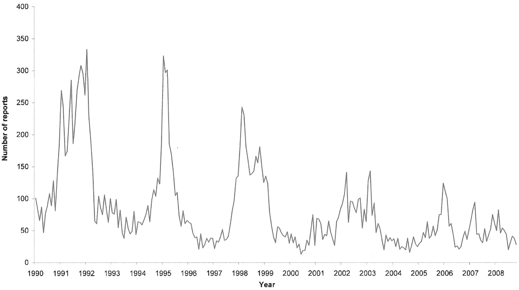
Laboratory reports to the Health Protection Agency Centre for Infections of infections due to Mycoplasma pneumoniae in England and Wales by date of report, 1990–2008 (4-weekly).
Chlamydophilapneumoniae
Epidemics occur in the community and in closed communities.93 94 95 [II] [II] [II] Its direct pathogenic role as a cause, as opposed to being associated with CAP, is not clear. The lack of a diagnostic gold standard means the frequency is unknown. Serological and PCR96 [Ib] results are highly variable between assays. Evidence that antibiotic therapy directed against this organism alters the course of the illness is lacking. When identified, other bacterial pathogens (eg, S pneumoniae) are often identified in the same host.97 98 99 [II] [II] [II] Patients may recover when antibiotics to which C pneumoniae is not sensitive are given.99 [II]
Chlamydophilapsittaci
Infection is acquired from birds and animals but human to human spread may occur. [II] Epidemics are reported in relation to infected sources at work (eg, poultry or duck workers). [II] Only 20% of UK cases have a history of bird contact.100 [II]
Coxiellaburnetii
Cases are most common in April to June, possibly related to the lambing and calving season. [II] Epidemics occur in relation to animal sources (usually sheep), but a history of occupational exposure is only present in 7.7% (95% CI 6.2% to 9.4%) of cases.101 [II]
Staphylococcusaureus
It is more common in the winter months. Coincident influenza-type symptoms are reported in 39% (95% CI 27% to 53%) of cases.6 35 36 102 [II] Evidence of coincident influenza virus infection is found in 39% (95% CI 17% to 64%) of those admitted to hospital,6 35 36 102 [II] and 50% (95% CI 25% to 75%) of those admitted to an ICU.33 34 42 103 [II]
Multiple case reports104 105 106 107 108 109 110 111 112 113 114 115 116 117 118 [III] and series of 2–11 patients,119 120 121 122 123 124 [II] both from the UK and worldwide, describe episodes of CAP caused by S aureus (either methicillin-sensitive S aureus (MSSA) or MRSA) capable of production of the Panton-Valentine Leucocidin toxin. Severe illness—with high mortality, bilateral lung shadowing and frequent lung cavitation—is common to these reports. No prospective studies have been performed to identify the true frequency of CAP due to this organism, but it appears to be rare at present.
Influenza virus
Annual epidemics of varying size are seen during the winter months.125 [II] Pneumonia complicates 2.9% (95% CI 1.4% to 5.4%) of cases in the community.126 [Ib] The frequency of staphylococcal pneumonia in patients with influenza symptoms is not known. Of adults with CAP admitted to UK hospitals in whom influenza infection is confirmed, 10% (95% CI 4.1% to 19.5%) have coincident S aureus infection. [II] Of those admitted to an ICU, the corresponding figure is 67% (95% CI 35% to 90%).34 42 43 103 [II]
Summary
The low frequency of legionella, staphylococcal, C psittaci and C burnetii infection in patients with CAP in both the community and in hospital, together with the likely high frequency of the relevant risk factors (outlined above) in the general population suggests that routine enquiry about such factors is likely to be misleading. [IV]
Only in those with severe illness where the frequency of legionella and staphylococcal infection is higher may enquiry about foreign travel and influenza symptoms be of predictive value. [IV]
Knowledge of increased mycoplasma activity in the community during an epidemic period may help guide the clinician to the increased likelihood of mycoplasma infection. [IV]
Section 4 Clinical features
4.1 Can the aetiology of CAP be predicted from clinical features?
There have been a large number of publications looking at the possibility of predicting the aetiological agent from the clinical features at presentation; however, while certain symptoms and signs are more common with specific pathogens, none allow accurate differentiation.127 128 [II] This led to a suggestion that the term “atypical” pneumonia be abandoned.128 As explained in Section 1.3.2, the term “atypical pathogens” remains useful and there is evidence that pleuritic pain is less likely in pneumonia secondary to these agents.129
Summary
The likely aetiological agent causing CAP cannot be accurately predicted from clinical features. [II]
The term “atypical” pneumonia should be abandoned as it incorrectly implies that there is a characteristic clinical presentation for patients with infection caused by “atypical” pathogens. [II]
4.2 Specific clinical features of particular respiratory pathogens
Clinical features associated with specific pathogens are described below and summarised in box 1.
Box 1 Some clinical features reported to be more common with specific pathogens (references are given in the text)
Streptococcus pneumoniae: increasing age, comorbidity, acute onset, high fever and pleuritic chest pain.
Bacteraemic S pneumoniae: female sex, excess alcohol, diabetes mellitus, chronic obstructive pulmonary disease, dry cough.
Legionella pneumophila: younger patients, smokers, absence of comorbidity, diarrhoea, neurological symptoms, more severe infection and evidence of multisystem involvement (eg, abnormal liver function tests, elevated serum creatine kinase).
Mycoplasma pneumoniae: younger patients, prior antibiotics, less multisystem involvement.
Chlamydophila pneumoniae: longer duration of symptoms before hospital admission, headache.
Coxiella burnetii: males, dry cough, high fever.
Streptococcuspneumoniae
One study using discriminant function analysis found pneumococcal aetiology to be more likely in the presence of cardiovascular comorbidity, an acute onset, pleuritic chest pain and less likely if patients had a cough or flu-like symptoms or had received an antibiotic before admission.130 [III]
Bacteraemic pneumococcal pneumonia was found to be more likely in those patients who had at least one of the following features: female, history of no cough or a non-productive cough, history of excess alcohol, diabetes mellitus or COPD.76 [II]
In high severity CAP where patients were admitted to an ICU, clinical features had little value in predicting the aetiological agent with the exception of those patients with fever (>39°C) or chest pain who were statistically more likely to have pneumococcal pneumonia.44 [II]
Legionellapneumophila
A variety of clinical features have been found to be more common in patients with legionella pneumonia, and yet most agree that it remains impossible to accurately differentiate on clinical grounds.131 132 Studies have reported L pneumophila to be more common in men,133 in young patients with lower rates of comorbid illness,134 in smokers135 and in those who have already received antibiotic therapy.133 135 Clinical features which might point towards L pneumophila as an aetiological agent include encephalopathy and other neurological symptoms, gastrointestinal symptoms, more severe infection, elevated liver enzymes, elevated creatine kinase and relatively less frequent upper respiratory tract symptoms, pleuritic chest pain and purulent sputum.82 133 136
Mycoplasmapneumoniae
One study has compared CAP due to M pneumoniae to patients with pneumococcal or legionella pneumonia. It reported that patients with mycoplasma pneumonia were younger, less likely to have multisystem involvement and more likely to have received an antibiotic before admission.23 By contrast, another report found no distinctive clinical features in patients with confirmed M pneumoniae pneumonia.137 [II]
Chlamydophilapneumoniae
A comparative study of patients with C pneumoniae and S pneumoniae pneumonia found the former more likely to present with headaches and a longer duration of symptoms before hospital admission.95 [II] A study from Israel reported no distinguishing clinical features for chlamydial pneumonia, except that it affected older patients than pneumococcal and mycoplasma infections.98 [II] A comparison of C pneumoniae and M pneumoniae confirmed the age difference between the groups and stated that, although clinical features could not be used to distinguish between the two, cough, hoarseness and rhinitis were all more common in M pneumoniae pneumonia.138 [III] Where C pneumoniae was the only pathogen identified, the illness was generally mild with non-specific symptoms.139 [II]
Coxiellaburnetii
CAP due to C burnetii (Q fever) causes non-specific clinical features.140 141 [II] Two reviews of Q fever have reported that infection was more common in younger men and that patients tended to present with dry cough and high fever.142 143 [III] Epidemiological features are discussed in Section 3.
Klebsiellapneumoniae
When compared with S pneumoniae, K pneumoniae was found to affect men more commonly and to present with a lower platelet count and leucopenia. Alcoholics were at particular risk of bacteraemic and fatal Klebsiella pneumonia.144 [III]
Some rarer community respiratory pathogens
CAP caused by Acinetobacter is seen more often in older patients with a history of alcoholism and has a high mortality.145 [III]
CAP due to Streptococcus milleri may indicate a dental or abdominal source of infection.146 [III] CAP due to viridans streptococci is associated with aspiration.147 [III]
4.3 CAP in elderly patients: are risk factors and clinical features different?
The classic symptoms and signs of pneumonia are less likely in elderly patients and non-specific features, especially confusion, are more likely.73 148 149 150 [II] Comorbid illness occurs more frequently in older patients with CAP and two studies have found the absence of fever to be more common than in younger patients with CAP.128 151 [II]
There is a high incidence of aspiration in elderly patients who present with CAP compared with controls (71% versus 10%).152 Case-controlled studies of pneumonia acquired in nursing homes have shown that both aspiration and pre-existing comorbid illnesses were more common in nursing home-acquired pneumonia than in others with CAP.77 148 [II] The in-patient mortality rate for nursing home-acquired pneumonia was higher than that for age matched patients with non-nursing home-acquired pneumonia.153 [II] The relationship between aetiology of CAP and the age of the patient is discussed in Section 3.
Summary
Elderly patients with CAP more frequently present with non-specific symptoms and have comorbid disease and a higher mortality, and are less likely to have a fever than younger patients. [II]
Aspiration is a risk factor for CAP in elderly patients, particularly nursing home residents. [II]
4.4 Aspiration pneumonia
Aspiration pneumonia embodies the concept of an infectious pneumonic process consequent upon the aspiration of colonised oropharyngeal or gastric contents. However, in practice, such causal linkage is seldom verified. Instead, the term “aspiration pneumonia” is commonly applied to situations when a patient with risk factors for aspiration presents with pneumonia. These risk factors include altered level of consciousness, neurological disorders such as stroke, presence of dysphagia and gastric disorders such as gastro-oesophageal reflux. When a broad definition of aspiration pneumonia is applied to pneumonia study cohorts, up to 10% of patients admitted to hospital with CAP are identified as having aspiration pneumonia.154 [II] This is likely to be an overestimate of the incidence of true aspiration pneumonia.
Studies of the bacteriology of pneumonia in patients with risk factors for aspiration vary widely in relation to inclusion criteria, patient characteristics and microbiological techniques used.44 72 154 155 156 157 158 [III] [III] [II] [II] [II] [Ib] [Ib] In true community acquired aspiration pneumonia, multiple pathogens including anaerobes are likely.
Section 5 Radiological, general and microbiological investigations
5.1 When should a chest radiograph be performed in the community for patients presenting with suspected CAP?
In UK practice, most CAP is managed in primary care where access to rapid chest radiography is limited. In this setting, clinicians have to identify the 5–12% with CAP from the majority with acute non-pneumonic lower respiratory tract infections or other diagnoses.3 24 [Ib] This challenge is particularly difficult in the presence of comorbid illnesses such as left ventricular failure, chronic lung disease or COPD and in the elderly who frequently present with non-specific symptoms and an absence of chest signs.148 [II]
No individual clinical symptom or sign is useful in discriminating CAP from other acute lower respiratory tract infections,159 160 [Ia] and there is poor interobserver reliability in eliciting respiratory signs.161 [II]
Woodhead et al23 [II] found that 39% of adults treated with antibiotics for an acute lower respiratory tract infection associated with new focal signs on chest examination had evidence of CAP on chest radiograph compared with 2% of patients who did not have new focal chest signs. By contrast, Melbye et al162 [II] found that respiratory symptoms and signs were of only minor value in differentiating patients with radiographic pneumonia in a study of 71 patients suspected by their general practitioners of having CAP. The clinical findings reported by the general practitioners to be most suggestive to them of CAP (typical history of cough, fever, dyspnoea and chest pains and lung crackles on examination) had low predictive values; only a short duration of symptoms (<24 h) was of significant predictive value.
Various prediction rules have been published for the diagnosis of CAP, [II] but generally have shown the need for confirmatory radiographic evidence. Statistical modelling was used by Diehr et al163 [II] to predict the presence of CAP in 1819 adults presenting to hospital outpatients with acute cough, 2.6% of whom had CAP on the chest radiograph. The presence of fever (>37.8°C), raised respiratory rate (>25 breaths/min), sputum production throughout the day, myalgia and night sweats, and absence of sore throat and rhinorrhoea were the only clinical features that predicted CAP when included in a diagnostic rule which had 91% sensitivity and 40% specificity.
Conversely, a number of studies have suggested that CAP can be safely ruled out in the absence of abnormal vital signs.159 160 [Ia] One study compared 350 adults presenting with acute respiratory symptoms to outpatient clinics and the emergency department in California where CAP had been diagnosed on the chest radiograph with an equal number of age-matched controls. The age range of patients was 21–91 years, with an average age of 65 years. The presence of either abnormal vital signs (fever >38°C, tachycardia >100/min and tachypnoea >20/min) or an abnormal physical examination of the chest (crackles, decreased breath sounds, dullness to percussion, wheeze) identified patients with radiographically confirmed CAP with a sensitivity of 95%, negative predictive value of 92% and specificity of 56%.164 [II] These findings have not been validated in the UK. Despite the age range included in this study, the reduced incidence of classical features of pneumonia and fever with increasing age at presentation (see Section 4.4) should be borne in mind when applying these results to elderly patients.
In practice, general practitioners manage the vast majority of patients pragmatically at first presentation. The important decision in patients presenting with a lower respiratory tract infection, or suspected CAP, is deciding whether to use an antibiotic, which one and how ill the patient is. Labelling the illness as pneumonia is less important.165
Recommendations
It is not necessary to perform a chest radiograph in patients with suspected CAP unless:
The diagnosis is in doubt and a chest radiograph will help in a differential diagnosis and management of the acute illness. [D]
Progress following treatment for suspected CAP is not satisfactory at review. [D]
The patient is considered at risk of underlying lung pathology such as lung cancer (see Section 5.6). [D]
5.2 When should a chest radiograph be performed in hospital for patients presenting with suspected CAP?
A chest radiograph is the cornerstone to confirming a diagnosis of CAP. In patients ill enough to require hospital referral for suspected CAP, a chest radiograph is essential to establishing the diagnosis of CAP or an alternative diagnosis, and therefore in guiding management decisions.
Antibiotic treatment of patients with suspected CAP prior to, or without, confirmation by chest radiography potentially leads to inappropriate and excessive antibiotic use.
The committee felt that the Department of Health’s “4 hour from presentation to admission, transfer or discharge” target for patients admitted to emergency departments represented a practice standard that should apply to all patients presenting to hospital (via the emergency department or acute medical unit) with suspected CAP.15
Recommendation
All patients admitted to hospital with suspected CAP should have a chest radiograph performed as soon as possible to confirm or refute the diagnosis. [D] The objective of any service should be for the chest radiograph to be performed in time for antibiotics to be administrated within 4 h of presentation to hospital should the diagnosis of CAP be confirmed.
5.3 Are there characteristic features that enable the clinician to predict the likely pathogen from the chest radiograph?
There are no characteristic features on the chest radiograph in CAP that allow confident prediction of the causative organism.98 166 167 168 [III] The lower lobes are affected most commonly, regardless of aetiology.
Multilobe involvement169 [II] at presentation and pleural effusions were more likely at presentation in bacteraemic pneumococcal pneumonia than in non-bacteraemic pneumococcal pneumonia or legionella pneumonia. Homogenous shadowing was less common in mycoplasma pneumonia than in the other types. Lymphadenopathy was noted in some cases of mycoplasma infections but not in the other types of infection. CAP due to S aureus appears to be more likely to present with multilobar shadowing, cavitation, pneumatoceles or spontaneous pneumothorax.170 [III] K pneumoniae has been reported to produce chest radiograph changes with a predilection for upper lobes (especially the right).171 [II] A bulging interlobar fissure and abscess formation with cavitation have also been reported, although the former is probably just a reflection of an intense inflammatory reaction that can occur in any severe infection such as pneumonia due to S aureus.170 [III]
Summary
There are no characteristic features of the chest radiograph in CAP that allows a confident prediction of the likely pathogen. [II]
5.4 What is the role of CT lung scans in CAP?
There are few data on the role of high-resolution CT lung scans in CAP. A small study has reported that high-resolution CT scans may improve the accuracy of diagnosing CAP compared with chest radiography alone.172 [II] Similarly, CT lung scans have improved sensitivity compared with standard chest radiographs in patients with mycoplasma pneumonia.173 [II] CT lung scans may be useful in subjects where the diagnosis is in doubt174 [III] but, in general, there is little role for CT scanning in the usual investigation of CAP.
With regard to aetiology, one study has reported a difference in CT appearances in 18 patients with CAP due to bacterial infections compared with 14 patients with atypical pathogens.175 [III]
Summary
CT scanning currently has no routine role in the investigation of CAP. [II]
5.5 How quickly do chest radiographs improve after CAP?
Radiographic changes resolve relatively slowly after CAP and lag behind clinical recovery. In one study, complete resolution of chest radiographic changes occurred at 2 weeks after initial presentation in 51% of cases, in 64% by 4 weeks and 73% at 6 weeks.176 Clearance rates were slower in elderly patients, those with more than one lobe involved at presentation, smokers and inpatients rather than outpatients. Multivariate analysis showed that only age and multilobe involvement were independently related to rate of clearance. Age was also a major factor influencing rate of radiographic recovery in the BTS multicentre CAP study.6 [Ib] A study of patients over 70 years of age showed 35%, 60% and 84% radiographic resolution at 3, 6 and 12 weeks, respectively.177 [II] C-reactive protein (CRP) levels >200 mg/l were also linked to slower radiographic resolution.178 [III] When chest radiographs of patients with bacteraemic pneumococcal pneumonia were followed, only 13% had cleared at 2 weeks and 41% at 4 weeks.179 [III] Pneumonias caused by atypical pathogens clear more quickly. The clearance rate has been reported to be faster for mycoplasma pneumonia than for legionella or pneumococcal pneumonia, which may take 12 weeks or more.166 [III] In a series of patients with C burnetii pneumonia, 81% of the chest radiographs had returned to normal within 4 weeks.143 [III]
Radiographic deterioration after admission to hospital was more common with legionella (65% of cases) and bacteraemic pneumococcal pneumonia (52%) than with non-bacteraemic pneumococcal (26%) or mycoplasma pneumonia (25%).166 [III] Residual pulmonary shadowing was found in over 25% of cases of legionella and bacteraemic pneumococcal cases. Deterioration after admission has also been reported in over half of cases of S aureus pneumonia.170 [III] Radiographic deterioration after hospital admission appears to be commoner in older patients (aged ⩾65 years).151 [II]
Summary
Radiological resolution often lags behind clinical improvement from CAP, particularly following legionella and bacteraemic pneumococcal infection. [III]
Pneumonia caused by atypical pathogens clears more quickly than pneumonia caused by bacterial infection. [III]
Radiological resolution is slower in elderly patients and where there is multilobar involvement. [Ib]
5.6 When should the chest radiograph be repeated during recovery and what action should be taken if the radiograph has not returned to normal?
Repeat chest radiographs are probably often ordered unnecessarily following CAP.180 [IVa] Although it has become usual practice to repeat the chest radiograph on hospital discharge and again at “routine” hospital clinic follow-up at around 6 weeks later, there is no evidence on which to base a recommendation regarding the value of this practice in patients who have otherwise recovered satisfactorily.
The main concern is whether the CAP was a complication of an underlying condition such as lung cancer. This concern will depend on a variety of factors such as age, smoking status, pre-existing conditions such as COPD and the clinical condition of the patient. In a study of 236 adults presenting to their general practitioner with a clinical diagnosis of CAP, 10 were found to have underlying lung cancer on investigation. There was a high frequency of lung cancer in older smokers (6 of 36 (17%) smokers aged >60 years), suggesting that a chest radiograph was particularly indicated in this group of patients with CAP in the community.23 [II] Studies of CAP in hospital often exclude patients found to have lung cancer, making it difficult to assess how frequently lung cancer presents acutely with CAP. In one study of 162 adults hospitalised with suspected CAP, the diagnosis was accepted in only 127, 10 (6%) of the 162 being found to have cancer.36 [II] Another study found only 13 (1.3%) of 1011 patients hospitalised with CAP to have an underlying lung cancer on investigation.181 [III] Eight of these were detected on the admission chest radiograph and the others were detected because of unsatisfactory clinical recovery. They concluded that a convalescent radiograph was useful in detecting occult lung cancer only if signs or symptoms persisted after a month or so.
The practice of performing bronchoscopy in patients admitted to hospital with CAP prior to hospital discharge has been investigated.182 [III] In patients aged >50 years or who were current or ex-smokers, 14% were found to have an abnormality at bronchoscopy (11% had a bronchial carcinoma diagnosed).
Recommendations
The chest radiograph need not be repeated prior to hospital discharge in those who have made a satisfactory clinical recovery from CAP. [D]
A chest radiograph should be arranged after about 6 weeks for all those patients who have persistence of symptoms or physical signs or who are at higher risk of underlying malignancy (especially smokers and those aged >50 years) whether or not they have been admitted to hospital. [D]
Further investigations which may include bronchoscopy should be considered in patients with persisting signs, symptoms and radiological abnormalities at around 6 weeks after completing treatment. [D]
It is the responsibility of the hospital team to arrange the follow-up plan with the patient and the general practitioner for those patients admitted to hospital (see Section 7.5). [D]
5.7 What general investigations should be done in a patient with suspected CAP in the community?
General investigations are performed to assess severity (see Section 6), to assess the impact on or to detect the presence of any comorbid disease, to provide some pointer to the particular aetiological agent or group of pathogens, identify complications and to monitor progress (see Section 9).
It may be appropriate to perform investigations in selected patients, especially if there is delayed improvement on review. However, no firm recommendations can be offered. It is a matter of clinical judgement.
Recommendations
General investigations are not necessary for the majority of patients with CAP who are managed in the community. [C] Pulse oximeters allow for simple assessment of oxygenation. General practitioners, particularly those working in out-of-hours and emergency assessment centres, should consider their use (see Section 7.1). [D]
Pulse oximetry should be available in all locations where emergency oxygen is used. [D]
5.8 What general investigations should be done in patients admitted to hospital?
Apart from the chest radiograph essential for diagnosis, the only other simple non-microbiological tests that influence immediate management are the urea, which informs severity assessment, and oxygen saturation, which affects supportive management and track and trigger systems in accordance with the BTS guideline for emergency oxygen use in adult patients.183
In addition, it is normal practice to take blood for a full blood count, urea and electrolytes, liver function tests and CRP. These often help to identify important underlying or associated pathologies including renal or hepatic disease and haematological or metabolic abnormalities.
A white cell count of >15×109/l strongly implicates a bacterial (particularly pneumococcal) aetiology, although lower counts do not exclude a bacterial cause.184 [III] A white cell count of >20×109/l or <4×10⁹/l is an indicator of severity (see Section 6).
Considering the role of CRP in the diagnosis of CAP, a prospective study performed in Spain reported a 96% specificity for CAP using a threshold CRP level of >100 mg/l.185 [II] Criticisms of this study are the small number of patients in one group and the fact that patients with infective exacerbations of COPD were excluded. Another study showed that a raised CRP level on admission is a relatively more sensitive marker of pneumonia than an elevated temperature or raised white cell count. All patients with CAP had CRP levels >50 mg/l and 75% of patients had levels >100 mg/l.186 [II] In the same paper it was reported that a CRP level of >100 mg/l helped to distinguish CAP from acute exacerbations of COPD. Another group found that only 5% of patients admitted with CAP had CRP levels <50 mg/l.187 [III] Although not yet widely available, a bedside finger-prick CRP test has been used to predict CAP in 168 patients presenting with acute cough and, at a cut-off of 40 mg/l, was found to have a sensitivity of 70% and a specificity of 90% independent of any clinical characteristics.188 [II] CRP levels are generally higher in patients who have not received antibiotic therapy before admission.186 [II]
With regard to predicting the microbial aetiology of CAP, higher CRP levels have been associated with pneumococcal pneumonia (especially if complicated by bacteraemia) compared with mycoplasma or viral pneumonias,189 [III] and in legionella pneumonia compared with all other identified single aetiologies.190 [II]
Data relating to CRP as a marker of severity and of treatment failure are discussed in Sections 6.2.1 and 7.3, respectively).
There are a number of studies examining the role of other biomarkers in CAP such as procalcitonin, however these assays are not currently widely available.
Summary
The published evidence to date suggests that measurement of CRP on admission may be helpful in distinguishing pneumonia from other acute respiratory illnesses. [III]
Recommendations
All patients should have the following tests performed on admission:
Oxygenation saturations and, where necessary, arterial blood gases in accordance with the BTS guideline for emergency oxygen use in adult patients. [B+]
Chest radiograph to allow accurate diagnosis. [B+]
Urea and electrolytes to inform severity assessment. [B+]
CRP to aid diagnosis and as a baseline measure. [B+]
Full blood count. [B−]
Liver function tests. [D]
5.9 Why are microbiological investigations performed in patients with CAP?
Establishing the microbial cause of CAP is useful for several reasons:
Identification of pathogens and antibiotic sensitivity patterns permits selection of optimal antibiotic regimens. To date there has been a habit to continue broad-spectrum empirical antibiotics even if a specific pathogen has been identified. However, with the increasing problem of antibiotic resistance and HCAIs such as C difficile infection, the balance has now swung towards focusing down antibiotic therapy whenever possible.
Targeted and narrow-spectrum antibiotic therapy limits drug costs, the threat of antibiotic resistance and adverse drug reactions such as C difficile-associated diarrhoea.
Specific pathogens have public health or infection control significance, including legionella, psittacosis, C burnetii, influenza A and multiresistant organisms. Patients with these infections should be identified quickly so that appropriate treatment and control measures can be implemented.
Microbiological investigations allow monitoring of the spectrum of pathogens causing CAP over time. This allows trends regarding aetiology and antibiotic sensitivity to be tracked for public health needs.
Unfortunately, microbiological investigations are insensitive and often do not contribute to initial patient management.191 [III] In detailed prospective aetiology studies the microbial cause is not found in 25–60% of patients,23 192 [II] [II] and the yield is even lower in routine hospital practice.193 194 [III] [III] More recent studies including the use of PCR and antigen detection techniques have not generally increased the proportion of patients with a specific aetiological diagnosis.195 196 [II] [II] In contrast, one recent prospective study of 105 adults with CAP197 [II] comparing a multiplex real-time PCR for a range of “atypical pathogens” and respiratory viruses with conventional methods reported a microbiological diagnosis in 80 patients (76%) using the real-time PCR compared with 52 patients (49.5%) using conventional methods. However, no urine antigen testing for either legionella or pneumococcal infection was included in the study, and most of the increase in diagnostic yield obtained was due to enhanced detection of rhinoviruses and coronaviruses. Nevertheless, such studies point the way forward for improving aetiological diagnosis in CAP.
Several studies198 199 200 201 [II] [II] [II] [II] have examined the positivity rate of routine microbiological investigations (blood and sputum cultures) for patients with CAP. These studies provide further evidence that the overall sensitivity of such tests in CAP is low, particularly for patients with low severity CAP and no comorbid disease, and for those who have received antibiotic therapy prior to admission. One study200 demonstrated a direct correlation between the severity of pneumonia (using the Fine Pneumonia Severity Index (PSI)) and blood culture positivity rate, and questioned the value of routine blood cultures for patients in PSI risk classes I–III (ie, low severity). However, another study found poor correlation of blood culture positivity with the PSI among patients hospitalised with CAP.201 [II]
Recommendations
Microbiological tests should be performed on all patients with moderate and high severity CAP, the extent of investigation in these patients being guided by severity. [D]
For patients with low severity CAP the extent of microbiological investigations should be guided by clinical factors (age, comorbid illness, severity indicators), epidemiological factors and prior antibiotic therapy. [A−]
Where there is clear microbiological evidence of a specific pathogen, empirical antibiotics should be changed to the appropriate pathogen-focused agent unless there are legitimate concerns about dual pathogen infection. [D]
5.10 What microbiological investigations should be performed in patients with suspected CAP in the community?
Comments about the pros and cons of different microbiological investigations are given below in Section 5.11. Many of these investigations will not be appropriate for patients with CAP managed in the community. Such patients are not usually severely ill, are at low risk of death and delays in transport of specimens to the laboratory reduces the yield of bacterial pathogens (especially S pneumoniae) from sputum cultures. Results are often received too late by the general practitioner to be of much practical value in initial management.
Recommendations
For patients managed in the community, microbiological investigations are not recommended routinely. [D]
Examination of sputum should be considered for patients who do not respond to empirical antibiotic therapy. [D]
Examination of sputum for Mycobacterium tuberculosis should be considered for patients with a persistent productive cough, especially if malaise, weight loss or night sweats, or risk factors for tuberculosis (eg, ethnic origin, social deprivation, elderly) are present. [D]
Urine antigen investigations, PCR of upper (eg, nose and throat swabs) or lower (eg, sputum) respiratory tract samples or serological investigations may be considered during outbreaks (eg, Legionnaires’ disease) or epidemic mycoplasma years, or when there is a particular clinical or epidemiological reason. [D]
5.11 What microbiological investigations should be performed in patients admitted to hospital with CAP?
The investigations that are recommended for patients admitted to hospital are summarised in table 4. More extensive microbiological investigations are recommended only for patients with moderate or high severity CAP, unless there are particular clinical or epidemiological features that warrant further microbiological studies. Comments and recommendations regarding specific investigations are given below.
5.11.1 Blood cultures
Microbial causes of CAP that can be associated with bacteraemia include S pneumoniae, H influenzae, S aureus and K pneumoniae. Isolation of these bacteria from blood cultures in patients with CAP is highly specific in determining the microbial aetiology. Bacteraemia is also a marker of illness severity. However, many patients with CAP do not have an associated bacteraemia. Even in pneumococcal pneumonia the sensitivity of blood cultures is at most only 25%,94 202 [II] [II] and is even lower for patients given antibiotic treatment before admission.129 [II] Several predominantly retrospective North American studies and reviews203 204 205 206 [II] [III] [III] [III] have questioned the utility of routine blood cultures in patients hospitalised with CAP on grounds of low sensitivity, cost and negligible impact on antimicrobial management. However, despite these limitations, most continue to recommend blood cultures in high severity CAP.
Recommendations
Blood cultures are recommended for all patients with moderate and high severity CAP, preferably before antibiotic therapy is commenced. [D]
If a diagnosis of CAP has been definitely confirmed and a patient has low severity pneumonia with no comorbid disease, then blood cultures may be omitted. [A−]
5.11.2 Sputum cultures
Sputum cultures may identify the causative agent in CAP including unexpected or antibiotic-resistant pathogens such as S aureus or antimicrobial-resistant pneumococci. Routine sputum cultures are, however, neither very sensitive nor specific207 [Ia] and often do not contribute to initial patient management.208 [II]
Problems include:
The inability of patients to produce good specimens.
Prior exposure to antibiotics.
Delays in transport and processing.
Difficulty in interpretation due to contamination of the sample by upper respiratory tract flora, which may include potential pathogens such as S pneumoniae and “coliforms” (especially in patients already given antibiotics).
Recommendations
Sputum samples should be sent for culture and sensitivity tests from patients with CAP of moderate severity who are able to expectorate purulent samples and have not received prior antibiotic therapy. Specimens should be transported rapidly to the laboratory. [A−]
Culture of sputum or other lower respiratory tract samples should also be performed for all patients with high severity CAP or those who fail to improve. [A−]
Sputum cultures for Legionella spp should always be attempted for patients who are legionella urine antigen positive in order to provide isolates for epidemiological typing and comparison with isolates from putative environmental sources. [D]
5.11.3 Sputum Gram stain
The value of performing a Gram stain on expectorated sputum has been widely debated. A meta-analysis review concluded that the sensitivity and specificity of sputum Gram stain in patients with CAP varied substantially in different settings.209 [Ia] The presence of large numbers of Gram-positive diplococci in purulent samples from patients with CAP can indicate pneumococcal pneumonia.210 [II] A study of 1669 consecutive adult patients with CAP found that good quality sputum samples with a predominant bacterial morphotype on Gram stain (ie, the test was useful) were obtained from only 14.4% of patients overall and, while Gram-positive diplococci as the predominant morphotype was highly specific for S pneumoniae, no severity subgroup of patients (assessed using the PSI) could be identified in whom the test would be of greater utility.211 [III] A similar study212 [III] of 347 patients with CAP concluded that Gram stain of sputum was useful in guiding microbiological diagnosis in just 23% of patients and unreliable in patients who had received antimicrobial treatment prior to sample collection. There are many factors which need to be borne in mind when considering the reliability and usefulness of Gram stain results. These are summarised below:
Advantages
Quick and relatively inexpensive.
Can assess quality of samples (cytological content) with rejection of poor quality samples.
Can aid the interpretation of culture results and occasionally give an early indication of possible aetiology.
Disadvantages
Strict criteria for interpretation require appropriate operator training.
Validity of results is directly related to the experience of the interpreter.213 [II]
Sputum Gram stain correlates poorly with culture results in conditions other than CAP.214 [II] This poses practical difficulties for laboratories that frequently have to interpret results with little or no clinical information.
Lack of availability: a recent survey of diagnostic microbiology laboratories in England and Wales215 [III] revealed that, of 138 respondents, 53 laboratories (38%) do not provide a sputum Gram stain service at all and, of the remainder, 52 laboratories (38%) do so only on special request. Thus, ready availability of sputum Gram stain cannot be assumed. This lack of availability reflects the opinion of many microbiologists that sputum examination is rarely helpful in the diagnosis of CAP.
Recommendations
Clinicians should establish with local laboratories the availability or otherwise of sputum Gram stain. Where this is available, laboratories should offer a reliable Gram stain for patients with high severity CAP or complications as occasionally this can give an immediate indicator of the likely pathogen. Routine performance or reporting of sputum Gram stain on all patients is unnecessary but can aid the laboratory interpretations of culture results. [B−]
Samples from patients already in receipt of antimicrobials are rarely helpful in establishing a diagnosis. [B−]
Laboratories performing sputum Gram stains should adhere to strict and locally agreed criteria for interpretation and reporting of results. [B+]
5.11.4 Other tests for Streptococcuspneumoniae
Pneumococcal antigen detection
Pneumococcal antigens can be detected in various body fluids during active pneumococcal infection, including sputum, pleural fluid, serum and urine. Antigen detection is less affected by prior antibiotic therapy and the detection of antigenaemia has a correlation with clinical severity.216 [IVb]
A commercial immunochromatographic strip test (BINAX NOW) for detection of pneumococcal antigen in urine has been introduced in the last few years and been widely taken up. Numerous studies217 218 219 220 221 222 [II] [II] [II] [II] [II] [III] have evaluated positively the clinical and diagnostic utility and generally good sensitivity and specificity of the pneumococcal urine antigen test in the diagnosis of pneumococcal pneumonia in adults. The studies have shown the usefulness of this assay in determining the aetiology of CAP, with significantly greater sensitivity rates than routine blood or sputum cultures. In addition, the test remains positive in 80–90% of patients for up to 7 days after starting antimicrobial treatment,223 [II] and may also be applied to other relevant sample types such as pleural fluid.224 [III]
Pneumococcal PCR
Many polymerase chain reaction (PCR)-based methods for detection of pneumococcal DNA in clinical samples have been published, varying in precise methodology and the specific pneumococcal DNA target(s) sought. However, relatively few studies report comprehensive clinical—as opposed to analytical sensitivity—evaluations of pneumococcal PCRs in the diagnosis of CAP. One retrospective study225 [III] compared three different PCR methodologies for use on EDTA blood samples from 175 bacteraemic patients collected at hospital admission (95 pneumococcal bacteraemia and 80 with bacteraemia due to other organisms). The best sensitivity obtained was 45% versus the gold standard of a positive blood culture for S pneumoniae. The specificity of all three methods was good at 97–100%. The authors concluded that blood PCR offers no advantage over conventional blood culture for pneumococcal diagnosis in bacteraemic patients and is unlikely to be sufficiently sensitive for diagnosis of non-bacteraemic pneumococcal pneumonia. Pneumococcal PCR has also been applied to sputum and other respiratory tract samples. However, obtaining a good quality sputum sample, as described above, remains problematic and, more importantly, PCR is not readily able to distinguish colonisation from infection of the respiratory tract.226 [III] Pneumococcal PCR has little to offer for the diagnosis of CAP at this time, being insufficiently sensitive and specific for routine use.
Recommendations
Pneumococcal urine antigen tests should be performed for all patients with moderate or high severity CAP. [A−]
A rapid testing and reporting service for pneumococcal urine antigen should be available to all hospitals admitting patients with CAP. [B+]
5.11.5 Tests for Legionnaires’ disease
Legionella pneumonia can be severe and carries a significant mortality. Prompt diagnosis is important both for patient management and for public health investigations. Risk factors for legionella infection include recent travel (within 10 days of onset), certain occupations, recent repair to domestic plumbing systems and immunosuppression.
Urine antigen detection
Detection of L pneumophila urinary antigen by enzyme immunoassay (EIA) is established as a highly specific (>95%) and sensitive (∼80%) test227 [III] for the detection of infections caused by L pneumophila serogroup 1, the commonest cause of sporadic and travel CAP cases in the UK. Rapid results can be obtained at an early stage of the illness, and this is a valuable method in the early diagnosis of legionella infection.92 [III] It is now widely applied in high severity CAP. A recent survey of diagnostic microbiology laboratories in England and Wales215 [III] revealed that, of 138 respondents, 136 laboratories (99%) offered this test for patients with CAP.
One study looked at the value of rapid legionella urine antigen testing in a large outbreak of Legionnaires’ disease caused by L pneumophila serogroup 1 in Holland.228 [III] This showed a higher test positivity rate for patients with severe legionella infection. The authors also demonstrated that the results of rapid testing could be used to start early legionella appropriate antibiotic management resulting in an improved outcome, as shown by reducing both mortality and the need for intensive care. In another prospective study of sporadic CAP in adults, the early detection of urine legionella antigen positively influenced the management of seven of nine patients in whom it was detected.37 [Ib]
There are several commercial assays available, including a rapid immunochromatographic test. These assays principally detect infection with L pneumophila serogroup 1. They do not reliably detect antigen from other serogroups or legionella species which can cause infection in immunocompromised patients who may present with CAP, or recently hospitalised patients. This has particular significance in nosocomial infection. In one study of culture confirmed cases, while the sensitivity of commercially available urine antigen tests was 93.7% for travel-associated cases and 86.5% for community-acquired cases, it was only 45% for nosocomial cases.229 [III]
Legionella direct immunofluorescence tests
L pneumophila can be detected by direct immunofluorescence (DIF) on invasive respiratory samples such as bronchial aspirates. L pneumophila specific reagents should be used, and not hyperimmune rabbit antisera which are poorly specific. The value of performing DIF on expectorated sputum samples is less well established. A considerable degree of laboratory expertise is required for processing and interpretation and, in view of the widespread availability of urine antigen tests for legionella diagnosis, the use of DIF has declined in recent years in the UK.
Culture
The culture of legionella from clinical samples (principally respiratory samples, including sputum) is very important and every effort should be made to diagnose by this method. Culture is 100% specific and is the only reliable method of detecting infection with non-pneumophila legionella species. Culture is also valuable for epidemiological investigations, allowing phenotypic and genotypic comparison of clinical and environmental legionella strains.
Problems with culture include: the inability of many patients with legionella pneumonia to produce sputum samples; prior antibiotic therapy; laboratory time and cost in processing samples; and lack of rapid results (legionella cultures need to be incubated for up to 10 days). Few laboratories will set up legionella cultures on respiratory samples unless specifically requested to do so. Culture should always be attempted from urine antigen positive patients and in suspected nosocomial legionella infection.
Serology
The diagnosis by determination of antibody levels was the mainstay of diagnosis of legionella pneumonia in the past. Serological assays previously employed in the UK were highly specific, although false positive results due to a serological cross-reaction may occur in patients with recent Campylobacter infection.230 [II] Serological reagents for legionella diagnosis are no longer available from the Health Protection Agency. Their place has been filled by a number of commercially available serological assays of varying sensitivity and specificity. It is currently recommended that, where a diagnosis of legionella infection relies solely on the results of serological testing, the sample should be referred to a reference laboratory for confirmation.
PCR
Detection of legionella DNA by PCR from respiratory samples is still only available as a reference laboratory or research tool, although it is becoming more widely available.
Recommendations
Investigations for legionella pneumonia are recommended for all patients with high severity CAP, for other patients with specific risk factors and for all patients with CAP during outbreaks. [D]
Legionella urine antigen tests should be performed for all patients with high severity CAP. [B+]
A rapid testing and reporting service for legionella urine antigen should be available to all hospitals admitting patients with CAP. [B+]
As the culture of legionella is very important for clinical reasons and source identification, specimens of respiratory secretions, including sputum, should be sent from patients with high severity CAP or where Legionnaires’ disease is suspected on epidemiological or clinical grounds. [D] The clinician should specifically request legionella culture on laboratory request forms.
Legionella cultures should be routinely performed on invasive respiratory samples (eg, obtained by bronchoscopy) from patients with CAP. [D]
For all patients who are legionella urine antigen positive, clinicians should send respiratory specimens such as sputum and request legionella culture. [D] This is to aid outbreak and source investigation with the aim of preventing further cases.
5.11.6 Tests for Mycoplasmapneumoniae
The mainstay of conventional diagnosis at the present time is by serology, although diagnosis by specific PCR is likely to become increasingly available. Culture of M pneumoniae is generally not available in diagnostic laboratories.
The commonest serological assay used historically was the complement fixation test (CFT), but there are various alternative assays such as microparticle agglutination and EIAs. The CFT is still regarded as the “gold standard” to which other assays have been compared, although it does lack sensitivity and specificity. A comparison of various mycoplasma antibody assays (including IgM and CFT tests) concluded that there is no single assay with significantly better sensitivity and specificity than the others.231 [III] Elevated CFT titres are usually detected no earlier than 10–14 days after the onset of mycoplasma infection, but the insidious onset and slow progression of symptoms means that many patients admitted to hospital with mycoplasma CAP have elevated titres on or shortly after admission.
One study232 [II] compared 12 commercially available serodiagnostic assays for M pneumoniae with the CFT using serum samples from patients with PCR-confirmed M pneumoniae infection and known onset dates. There were wide variations between the tests in sensitivity and specificity, CFT being the most specific (97%) although not especially sensitive (65%). The authors concluded that there are currently few commercial serological assays for the detection of M pneumoniae infections with appropriate performances in terms of sensitivity and specificity, and that PCR is likely to become increasingly important in specific diagnosis. In one series of patients with CAP,197 [II] application of PCR to respiratory tract samples doubled the detection rate of M pneumoniae infection versus serological testing alone from 5 to 10 of the 105 patients studied. Another study233 [III] described the application of real-time PCR to acute phase serum samples from patients with serologically (CFT) diagnosed mycoplasma pneumonia. Serum samples from 15 of 29 patients (52%) were M pneumoniae PCR positive, suggesting that serum PCR as opposed to respiratory tract sample PCR—with inherent issues of specimen quality—is worthy of further consideration. PCR diagnosis is already available in some centres in the UK, will become increasingly available, and is likely to replace serodiagnosis in the longer term.
Recommendations
Where available, PCR of respiratory tract samples such as sputum should be the method of choice for the diagnosis of mycoplasma pneumonia. [D]
In the absence of a sputum or lower respiratory tract sample, and where mycoplasma pneumonia is suspected on clinical and epidemiological grounds, a throat swab for Mycoplasma pneumoniae PCR is recommended. [D]
Serology with the complement fixation test and a range of other assays is widely available, although considerable caution is required in interpretation of results. [C]
5.11.7 Tests for Chlamydophila species
Culture
It is not appropriate for routine diagnostic laboratories to attempt culture of Chlamydophila from respiratory samples from patients with CAP as special laboratory precautions are required. (C psittaci is a “category 3 pathogen” indicating a high-risk pathogen that may put laboratory staff at risk of serious illness if infected occupationally.) C pneumoniae is very difficult to grow in the laboratory—culture is slow, time-consuming, expensive and insensitive.
Antigen detection
Chlamydophila antigen can be detected in respiratory samples using DIF with species- and genus-specific monoclonal antibodies.234 [II] Species-specific reagents are not available for C psittaci, which is antigenically highly diverse. DIF requires expertise in slide preparation and reading, and is not widely available in diagnostic laboratories. C pneumoniae can also be detected by DIF on throat swabs, with a comparable sensitivity to sputum.235 However, antigen may be detected for several months after “acute” infection, making interpretation difficult.
Chlamydophila antigen can also be detected in respiratory samples by EIA with comparable sensitivity to PCR,236 but this approach requires further studies.
Serology
Various serological assays are used in the diagnosis of respiratory Chlamydophila infections. The CFT is available in some diagnostic serology laboratories. Micro-immunofluorescence (MIF) and whole-cell immunofluorescence (WHIF) are specialised reference tests. Several EIAs have been described, and at least one is commercially available in the UK. Each of these assays has advantages and disadvantages, and there are particular problems in the serological diagnosis of C pneumoniae infections.
The CFT uses a genus-specific antigen and is relatively sensitive and specific for diagnosing psittacosis. However in adults, most infections with C pneumoniae are re-infections and these generate only a weak or absent CFT response.
The MIF and WHIF tests require considerable experience to read and interpret. They can detect a species-specific response, although this may be delayed for 4–6 weeks, especially with C pneumoniae re-infections. They may also miss C psittaci infections, depending on the particular serovars included in the test, and there are conflicting reports regarding the accuracy of these tests in reliably distinguishing chlamydial species.237 [IVb]
A commercial EIA has been used with success,238 [III] but has not been shown to be significantly superior to CFT.
Molecular techniques
Amplification of Chlamydophila DNA by PCR using genus- or species-specific primers has been reported from a variety of respiratory samples. These molecular techniques for diagnosis of respiratory Chlamydophila infection are mostly confined to research/reference/specialist laboratories at the present time although, as with all molecular diagnostics, such tests are likely to become increasingly available, often in commercial kit formats.
Recommendations
Chlamydophila antigen and/or PCR detection tests should be available for invasive respiratory samples from patients with high severity CAP or where there is a strong suspicion of psittacosis. [D]
The complement fixation test remains the most suitable and practical serological assay for routine diagnosis of respiratory Chlamydophila infections. [B−] There is no currently available serological test that can reliably detect acute infection due to C pneumoniae.
5.11.8 PCR and serology for other respiratory pathogens
PCR tests for a range of respiratory viruses and some atypical pathogens are becoming increasingly available through virology departments across the UK. The ready availability and extent of these investigations varies between individual laboratories and departments, although they are expected to increase over the foreseeable future. Where available, PCR tests are an extremely useful addition to the diagnostic armamentarium and have the advantage of being rapid (relevant on occasions for both clinical and infection control purposes) and sensitive, and so are to be preferred over serological tests. Evidence of influenza and/or other respiratory virus infections as a cause of primary viral pneumonia or as a copathogen in bacterial CAP is best sought by PCR of nose and throat swabs submitted in virus transport medium.
At present there are no readily available alternatives to serology for some of the atypical pathogens and, according to clinical and epidemiological parameters, serological investigations may be appropriate.
Respiratory serology usually comprises antibody tests for the atypical pathogens (M pneumoniae, Chlamydophila spp, C burnetii), influenza A virus, influenza B virus, adenovirus, respiratory syncytial virus and L pneumophila. Fewer laboratories rely on CFTs, which are time consuming and inconvenient to perform and have poor sensitivity and specificity.
There is little value in testing single serum samples taken within 7 days of the onset of CAP. Such samples can be stored until the follow-up (convalescent) sample is taken (7−10 days later) and the paired samples tested in parallel.
However, raised antibody titres—particularly to L pneumophila or M pneumoniae—may be found in some patients on or soon after hospital admission, particularly if the onset of symptoms is more than 7 days prior to admission. It is thus important that the date of onset of symptoms is clearly indicated on all serological request forms so that serum samples taken more than 1 week into the illness can be tested immediately.
A suggested algorithm for performing serological investigations is shown in fig 6.

Suggested algorithm for serological testing for patients with high severity community acquired pneumonia (CAP).
Recommendations
Where PCR for respiratory viruses and atypical pathogens is readily available or obtainable locally, this is preferred to serological investigations. [D]
Where available, paired serology tests can be considered for patients with high severity CAP where no particular microbiological diagnosis has been made by other means (eg, culture, urine antigen, PCR) and who fail to improve, and/or where there are particular epidemiological risk factors. [D] The date of onset of symptoms should be clearly indicated on all serological request forms. [D]
Serological tests may be extended to all patients admitted to hospital with CAP during outbreaks and when needed for the purposes of surveillance. The criteria for performing serology tests in these circumstances should be agreed locally between clinicians, laboratories and public health. [D]
Section 6 Severity assessment
6.1 Why is severity assessment important?
CAP presents to physicians both in primary and secondary care as a wide spectrum of illness from mild and self-limiting to life-threatening and occasionally fatal disease. This breadth of illness severity is reflected in the variable mortality rates reported by studies of CAP in different clinical settings.
The decision regarding the most appropriate site of care, including whether hospitalisation of a patient with CAP is warranted, is the first and single most important decision in the overall management of CAP. It has consequences both on the level of treatment received by the patient as well as the overall costs of treatment.30 [III] This decision is best informed by an accurate assessment of the severity of illness at presentation and the likely prognosis. The recognition of patients at low risk of complications—and therefore suitable for treatment out of hospital—has the potential to reduce inappropriate hospitalisation and consequently inherent morbidity and costs.
When hospital admission is required, further management is also influenced by illness severity. This includes the extent of microbiological investigation, the choice of initial empirical antimicrobial agents, route of administration, duration of treatment and level of nursing and medical care. Early identification of patients at high risk of death allows initiation of appropriate antibiotic therapy and admission to an intensive care setting where assisted ventilation can be readily initiated if necessary.
6.2 What clinical factors and investigations are associated with a poor prognosis on univariate and multivariate analysis?
A large number of studies conducted in hospital and intensive care settings have employed univariate analysis to identify risk factors associated with a poor prognosis. In-hospital mortality has been the most common outcome measure. Some studies have used admission to the ICU as the main outcome measure.239 [Ib] However, differences in ICU admission criteria make it difficult to compare results from these studies. This is reflected in the widely varying rates of admission of patients with CAP to ICU reported, ranging from 1–3% in New Zealand to 5% in the UK, 12–18% in the USA and 35% in Germany.5 6 239 240 241 242 [Ib] [Ib] [III] [II] [Ib] [III] In this document we have concentrated only on studies that have used mortality as the main outcome measure.
Univariate studies have suggested that over 40 different parameters are associated with mortality. An independent association of only a few of these risk factors with mortality has been consistently demonstrated by studies employing multivariate analysis. In particular, the following features are associated with an increased risk of mortality: increasing age;5 6 241 243 244 245 246 247 [Ib] [Ib] [Ib] [Ia] [Ib] [Ib] [III] [Ib] the presence of co-existing illnesses;5 243 244 245 247 248 249 250 251 [Ib] [III] [Ib] [Ib] [Ib] [III] [II] [III] [III] raised respiratory rate;5 6 129 241 242 243 251 252 253 254 [Ib] [Ib] [Ib] [Ib] [III] [Ia] [Ib] [Ib] [II] [III] mental confusion;6 129 244 246 253 255 [Ib] [Ib] [III] [III] [II] [Ib] low blood pressure;6 69 127 129 241 243 246 249 251 253 256 257 [Ib] [II] [III] [Ib] [Ib] [Ia] [III] [II] [III] [II] [II] [Ib] hypoxaemia;33 241 [II] [Ib] respiratory failure;5 33 44 258 [Ib] [II] [II] [Ib] high or low white cell count;6 243 [Ib] [Ia] bilateral or progressive chest radiograph changes;5 33 129 187 243 253 256 258 259 [Ib] [II] [Ib] [III] [Ia] [II] [II] [III,Ib] [Ib] and a positive blood culture.5 33 36 44 243 250 258 260 [Ib] [II] [II] [II] [Ia] [III] [Ib] [III]
The lack of pyrexia, which is easily overlooked in clinical practice, has been identified as an important poor prognostic factor in some studies of elderly patients.69 129 253 [II] [Ib] [II] Being previously bed-ridden and having been admitted from a nursing home are other poor prognostic factors that may be inter-related.5 129 153 247 [Ib] [Ib] [II] [Ib]
6.2.1 Biomarkers and inflammatory markers
A number of biomarker tests have been demonstrated on univariate and multivariate analyses to be independent prognostic factors for either 30-day or in-hospital mortality. These include procalcitonin,261 262 263 264 265 [II] [II] [Ib] [Ib] [II] triggering receptor expressed on myeloid cells-1 (TREM-1),266 [Ib] CD14,267 [Ib] pro-adrenomedullin,268 [Ib] CRP,257 269 [Ib] [II] pro-atrial natriuretic peptide270 271 [Ib] [III] and pro-vasopressin.270 [Ib] Further validation studies are required in most instances. Furthermore, determination of the majority of these biomarkers is not widely or routinely available at present.
Of the commonly available tests, one study has reported an association of a low CRP level of <100 mg/l at the time of hospital admission with reduced risk for 30-day mortality, need for mechanical ventilation and/or inotropic support, and complicated pneumonia.257 [Ib] This observation warrants further evaluation as a number of other studies have not found an association of admission CRP level with prognosis.261 264 265 272 [II] [Ib] [II] [Ib]
The level of D-dimers on admission has also been reported to be an independent prognostic marker.273 274 [Ib] [II] However, the clinical value of D-dimers over and above a clinical predictive model has not been established. Moreover, since D-dimers are mainly used in the diagnostic investigation of patients with suspected venous thromboembolism, introducing D-dimers solely as a prognostic marker in CAP will potentially lead to increased diagnostic uncertainty or confusion which, in turn, may result in delayed diagnosis or inappropriate management.
6.3 What predictive models for assessing severity on or shortly after hospital admission have been tested?
Clinical assessment of disease severity is dependent on the experience of the attending clinician, but such clinical judgement has been shown to result in apparent underestimation of severity.241 [Ib] No single prognostic factor of mortality is adequately specific and sensitive, so various severity scoring systems and predictive models have been developed in an attempt to help the clinician identify patients with pneumonia and a poor prognosis at an early stage.
However, predictive models have recognised limitations. First, no predictive model enables the unequivocal categorisation of patients into definite risk groups, and it is unrealistic to expect this.275 276 [IVb] [IVb] A predictive model is therefore not a substitute for clinical judgement, but should be considered as an adjunct to clinical judgement. Second, the value of severity-based practice guidelines in improving clinical outcomes has not yet been fully established.103 277 278 [III] [II] [II] Data demonstrating that severity-based practice guidelines are useful in increasing the proportion of patients managed as ambulatory outpatients without increasing morbidity or mortality have come from a cluster randomised trial, two large prospective observational studies and before-after implementation studies.279 280 281 282 [II] [Ib] [II] [III] Whether these benefits were mainly due to the use of a predictive model and therefore better stratification of patients to different management strategies, or simply due to better implementation of practice guidelines for the management of CAP283 [II] is difficult to differentiate. These encouraging results need confirmation in a wider setting, including the UK.
Conclusions regarding predictive models
No predictive model allows the unequivocal categorisation of patients into definite risk groups.
Predictive models based on severity are best viewed as useful adjuncts to clinical judgement.
Regular reassessment of severity during the course of hospital stay is mandatory if treatment is to be adjusted appropriately, avoiding the morbidity of overtreatment as well as the complications of undertreatment.
6.3.1 Pneumonia-specific predictive models
Pneumonia Severity Index (PSI)
The most widely studied predictive model in the management of CAP is the PSI developed in the USA.251 [Ib] The PSI is based on 20 variables that are used to derive a score which enables patients to be stratified into five risk categories based on 30-day mortality. It was developed to identify patients at low risk of mortality who might be suitable for ambulatory outpatient care, and it is in this setting that it is best validated. In the original study, mortality rates were 0.1%, 0.6%, 0.9%, 9.3% and 27% for risk classes I, II, III, IV and V, respectively. The PSI has been studied in over 50 000 patients worldwide and performs well. Patients in risk classes I–III are usually at low risk of mortality and therefore suitable for outpatient management. Due to the emphasis on age in the PSI, underestimation of severe pneumonia occasionally results. One study reported that up to 40% of patients with CAP who were hospitalised were assigned to low risk classes based on the PSI,284 [II] and another reported that ICU admission occurred in 27% of patients assigned to PSI risk classes I–III.285 [II] These reports reflect the importance of clinical judgment in assessing disease severity.
In clinical practice, the major limitation of the PSI with regard to its widespread and routine adoption in primary care, emergency departments or medical admission units is the complexity involved in the calculation of the score.
CURB65 score
The CURB65 score was developed based on a study of over 1000 prospectively studied patients with CAP from three countries: the UK, New Zealand and the Netherlands.255 [Ib] The 6-point CURB65 score, one point for each of Confusion, Urea >7 mmol/l, Respiratory rate ⩾30/min, low systolic (<90 mm Hg) or diastolic (⩽60 mm Hg) Blood pressure, age ⩾65 years (CURB65 score) based on information available at initial hospital assessment, enabled patients to be stratified according to increasing risk of mortality (score 0, 0.7%; score 1, 2.1%; score 2, 9.2%; scores 3–5, 15–40%). A similar pattern of increasing disease severity was reported when only clinical parameters were considered (CRB65) giving a 5-point score (risk of mortality for each score: score 0, 1.2%; score 1, 5.3%; score 2, 12.2%; scores 3–4, up to 33%).
Since then, the CURB65 score has been studied in over 12 000 patients.257 266 286 287 288 289 290 291 292 293 294 295 [Ib] [Ib] [II] [Ib] [Ib] [II] [Ib] [II] [III] [II] [Ib] [III] The results from all but one of the prospective validation studies indicate that the PSI and the CURB65 score perform equally well at discriminating patients into mortality risk groups. The CRB65 score has been specifically studied in over 6000 patients representing a mix of patients seen both in the community and in hospitals.255 286 287 289 296 297 298 299 [Ib] [II] [Ib] [II] [Ib] [II] [Ib] [II] All studies reported findings similar to the derivation study and, in certain studies, the CRB65 score was reported to be of similar discriminatory value to the CURB65 score.286 298 [II] [Ib] In prospective studies reporting from the UK, 41–45% of patients hospitalised with CAP had CURB65 scores of 0–1, 25–28% had a CURB65 score of 2 and 29–34% had CURB65 scores of 3 or more.255 257
Other predictive models have been developed, including models to predict admission to intensive care as the primary outcome, or to predict a composite outcome comprising either mortality or need for intensive care.246 287 291 294 300 301 302 303 [III] [Ib] [Ib] [Ib] [II] [III] [Ib] [Ib] Widespread validation of most of these models in different settings or patient cohorts has not yet occurred. Differences between studies in relation to criteria for intensive care admission need to be taken into account when interpreting these data.
6.3.2 Generic predictive models
Generic predictive models such as the sepsis score or APACHE II scoring system have also been tested in patients with CAP and been shown to predict outcome.189 258 297 304 [Ib] [IIb] [II] [IIb] However, general adoption of scoring systems originally designed for use within an intensive care setting is difficult, time-consuming and likely to be impractical. In addition, pneumonia-specific predictive models have been shown to outperform generic predictive models such as a modified Early Warning Score, in the context of CAP.248 289 [III] [II]
6.4 What severity assessment strategy is recommended for CAP?
We have been keen to recommend one severity assessment strategy that is applicable to adults of all ages, simple to remember and practical to implement both in the community and in hospital.
With these principles in mind, the CURB65 score in conjunction with clinical judgement is recommended as the initial severity assessment strategy in hospitals for CAP. The evidence base for the CURB65 score is robust and continues to increase, adding to the strength of the current recommendation. The simplified CRB65 score which only relies on clinical factors in conjunction with clinical judgement is recommended as the severity assessment strategy in community or primary care settings for CAP.
Summary of the CURB65 score
Confusion: New mental confusion, defined as an Abbreviated Mental Test score191 [II] (summarised in box 2) of 8 or less.
Urea: Raised >7 mmol/l (for patients being seen in hospital).
Respiratory rate: Raised ⩾30/min.
Blood pressure: Low blood pressure (systolic <90 mm Hg and/or diastolic ⩽60 mm Hg).
Age ⩾65 years.
Box 2 Abbreviated Mental Test
The Abbreviated Mental Test (each question scores 1 mark, total 10 marks)
Age
Date of birth
Time (to nearest hour)
Year
Hospital name
Recognition of two persons (eg, doctor, nurse)
Recall address (eg, 42 West Street)
Date of First World War
Name of monarchs
Count backwards 20 → 1
A score of 8 or less has been used to define mental confusion in the CURB65 severity score.
6.4.1 Clinical judgement
Clinical judgement is essential when deciding on the management of all patients with CAP, particularly in the following three areas with regard to severity assessment:
Interpretation of CURB65 score. Each patient must be managed individually and the interpretation of the CURB65 score is best refined through clinical judgement that takes into account all the clinical information available at the time. For instance, the combination of age <50 years, the absence of coexisting disease and a CRB65 or CURB65 score of 0 identifies patients with a very good prognosis who should be suitable for home treatment. In comparison, young patients with a respiratory rate of >40/min may warrant hospital supervised management despite a CRB65 or CURB65 score of 1. Clinical judgement is especially important in patients at high risk of death (CURB65 scores 3, 4 and 5) in whom decisions regarding intravenous administration of antibiotics or transfer to critical care facilities need to be made.
Stability of comorbid illnesses. The CURB65 score is primarily a CAP severity assessment tool. Pneumonia may result in a worsening of comorbid illness that warrants hospital or critical care management irrespective of severity of pneumonia.
Social circumstances. The morbidity associated with CAP negatively influences the functional status of patients and therefore the extent that patients may be able to manage at home within a given set of social circumstances. Patient choice should also be considered when determining social circumstances and the feasibility of further management at home.
Recommendations
Clinical judgement is essential in disease severity assessment. [D]
The stability of any comorbid illness and a patient’s social circumstances should be considered when assessing disease severity. [D]
6.5 Severity assessment of CAP in patients seen in the community
The severity assessment of CAP in patients seen in the community is shown in fig 7.

Severity assessment of community acquired pneumonia (CAP) in patients seen in the community (CRB65 severity score plus clinical judgement). DBP, diastolic blood pressure; SBP, systolic blood pressure.
Recommendations
For all patients, clinical judgement supported by the CRB65 score should be applied when deciding whether to treat at home or refer to hospital. [D]
Patients who have a CRB65 score of 0 are at low risk of death and do not normally require hospitalisation for clinical reasons. [B+]
Patients who have a CRB65 score of 1 or 2 are at increased risk of death, particularly with a score of 2, and hospital referral and assessment should be considered. [B+]
Patients who have a CRB65 score of 3 or more are at high risk of death and require urgent hospital admission. [B+]
When deciding on home treatment, the patient’s social circumstances and wishes must be taken into account in all instances. [D]
6.6 Severity assessment of CAP in patients seen in hospital
The severity assessment of CAP in patients seen in hospital is shown in fig 8.
Recommendations
For all patients, the CURB65 score should be interpreted in conjunction with clinical judgement. [D]
Patients who have a CURB65 score of 3 or more are at high risk of death. These patients should be reviewed by a senior physician at the earliest opportunity to refine disease severity assessment and should usually be managed as having high severity pneumonia. Patients with CURB65 scores of 4 and 5 should be assessed with specific consideration to the need for transfer to a critical care unit (high dependency unit or intensive care unit). [B+]
Patients who have a CURB65 score of 2 are at moderate risk of death. They should be considered for short-stay inpatient treatment or hospital-supervised outpatient treatment. [B+]
Patients who have a CURB65 score of 0 or 1 are at low risk of death. These patients may be suitable for treatment at home. [B+]
When deciding on home treatment, the patient’s social circumstances and wishes must be taken into account in all instances. [D]
6.7 Reviewing severity status after initial assessment in hospital
Summary
Regular and structured clinical review and reassessment of disease severity facilitates the stepping down and stepping up of antibiotic management. [Ib]
Recommendations
Regular assessment of disease severity is recommended for all patients following hospital admission. The “post take” round by a senior doctor and the medical team provides one early opportunity for this review. [D]
All patients deemed at high risk of death on admission to hospital should be reviewed medically at least 12-hourly until shown to be improving. [D]
Section 7 General management in the community and in hospital
7.1 What general management strategy should be offered to patients treated in the community?
Patients with CAP may present with fever, cough, sputum production or pleuritic pain and usually have localised signs on chest examination. They should be advised to rest and avoid smoking305 [IIb] and, especially when febrile, be encouraged to drink plenty of fluids. It is important to relieve pleuritic pain using simple analgesia such as paracetamol or non-steroidal anti-inflammatory drugs. Physiotherapy is of no proven benefit in acute pneumonia.306 [III] Nutritional status appears important both to the outcome and the risk of acquiring pneumonia and, in prolonged illness, nutritional supplements may be helpful. Patients with pneumonia are often catabolic and those aged >55 years who are malnourished appear to be at greater risk of developing pneumonia.150 307 [III] [III]
Patients with pneumonia often become hypoxic because pulmonary blood flow takes place through unventilated lung tissue. The clinical signs of hypoxia are non-specific and often difficult to recognise in the early stages. They include altered mental state, dyspnoea and tachypnoea. Respiratory rate should therefore always be assessed. Central cyanosis is unreliable both as a clinical sign and also as an indicator of tissue hypoxia. In contrast, pulse oximetry which measures arterial oxygen saturation (Spo2) is, in most situations, a simple and reliable method of assessing oxygenation. However, poor peripheral perfusion, jaundice and pigmented skin can produce a falsely low saturation and carboxyhaemoglobin a falsely high saturation. Pulse oximetry is now widely available in North America. A survey of 944 outpatients and 1332 inpatients with evidence of CAP enrolled from five sites in the USA and Canada reported increasing assessment of arterial oxygen saturation with pulse oximetry in up to 58% of outpatients and 85% of inpatients.308 [II] It is recommended that pulse oximetry, with appropriate training, should become more widely available in general practice for use in the assessment of patients who may have pneumonia and other acute respiratory illnesses. Oxygen saturation below 94% in a patient with CAP is an adverse prognostic feature and also an indication for oxygen therapy,309 [IVb] which will usually require urgent referral to hospital.
Patients who fall outside the low severity criteria for CAP should be assessed for the need for hospital referral (see Section 6). Social factors will also play an important part in the decision to refer a patient to hospital. Patients with moderate or high severity pneumonia should be admitted to hospital and managed, where possible, with input from a physician with an interest in respiratory medicine.
Recommendations
Patients with suspected CAP should be advised to rest, to drink plenty of fluids and not to smoke. [D]
Pleuritic pain should be relieved using simple analgesia such as paracetamol. [D]
The need for hospital referral should be assessed using the criteria recommended in section 6. [C]
Pulse oximetry, with appropriate training, should be available to general practitioners and others responsible for the assessment of patients in the out-of-hours setting for the assessment of severity and oxygen requirement in patients with CAP and other acute respiratory illnesses. [D]
7.2 What review policy should be adopted in patients managed in the community?
When to review a patient with CAP in the community will be determined by the initial severity assessment and other factors such as reliable help in the home. Patients assessed as being at low severity should improve on appropriate therapy within 48 h, at which time severity reassessment is recommended. Those who fail to improve within 48 h should be considered for hospital admission. Patients who do not fulfil the criteria for low severity and are being managed at home will require more frequent review.
Recommendations
Review of patients in the community with CAP is recommended after 48 h or earlier if clinically indicated. Disease severity assessment should form part of the clinical review. [D]
Those who fail to improve after 48 h of treatment should be considered for hospital admission or chest radiography. [D]
7.3 What general management strategy should be offered to patients in hospital?
Initial management
There is some evidence that use of a critical care pathway for patients referred to hospital can reduce the hospital admission of low-risk patients and can also rationalise inpatient management.310 [Ib]
All patients referred to hospital with CAP should have a chest radiograph (if not already performed in the community) and should have oxygenation assessed by pulse oximetry, preferably while breathing air. Those with Spo2 <94% should have arterial blood gas measurements, as should all patients with features of high severity pneumonia. Knowledge of the inspired oxygen concentration is essential to the interpretation of blood gas measurements and should be clearly recorded with the blood gas result.
Continuous oxygen therapy is usually indicated for those patients with arterial oxygen tension (Pao2) <8 kPa.183 The aim of oxygen therapy should be to maintain Pao2 ⩾8 kPa or Spo2 94–98% in patients who are not at risk of hypercapnic respiratory failure. In nearly all cases of CAP, unless complicated by severe COPD with ventilatory failure, high concentrations of oxygen of 35% or more are indicated and can be safely used.
High concentration oxygen therapy given to patients at risk of hypercapnic respiratory failure, such as patients with moderate or severe COPD, can reduce hypoxic drive and increase ventilation-perfusion mismatching. In such patients, initial treatment should be with low oxygen concentrations (24–28%), guided by arterial blood gas measurements, with the aim of keeping oxygen saturation at 88–92% without causing a fall in arterial pH (pH <7.35 or [H+] >45 nmol/l.183 In selected cases, non-invasive ventilation or respiratory stimulants may be of value and transfer to a high dependency unit or ICU area should be considered. If the patient is hypercapnic (Paco2 >6 kPa or 45 mm Hg) and acidotic (pH <7.35 or [H+] >45 nmol/l), ventilatory support should be considered, especially if acidosis has persisted for more than 30 min despite appropriate therapy.
Patients admitted with pneumonia should be assessed for volume depletion and may require intravenous fluids.
The use of unfractionated or low molecular weight heparins for the prevention of venous thromboembolism in patients with respiratory tract infections has been demonstrated in clinical trials.311 312 313 [Ia]
A randomised study (n = 458) found early mobilisation (sitting out of bed for at least 20 min within the first 24 h of hospital admission with increasing mobility on subsequent days) decreased length of stay by 1.1 days.314 [Ib] Further studies are needed.
Routine airway clearance has not been shown to improve outcome.315 [Ib]
While chest physiotherapy is of no proven value in acute pneumonia,315 [III] a single randomised trial using positive expiratory pressure has suggested that “bottle-blowing” into an underwater seal bottle on 10 occasions daily can shorten hospital stay.316 [II]
In cases of severe pneumonia requiring prolonged hospital admission, increased nutritional support (whether enteral, parenteral or via nasogastric feeding) should be arranged.
Recommendations
All patients should receive appropriate oxygen therapy with monitoring of oxygen saturations and inspired oxygen concentration with the aim to maintain Pao2 at ⩾8 kPa and Spo2 94–98%. High concentrations of oxygen can safely be given in patients who are not at risk of hypercapnic respiratory failure. [D]
Oxygen therapy in patients at risk of hypercapnic respiratory failure complicated by ventilatory failure should be guided by repeated arterial blood gas measurements. [C]
Patients should be assessed for volume depletion and may require intravenous fluids. [C]
Prophylaxis of venous thromboembolism with low molecular weight heparins should be considered for all patients who are not fully mobile. [A+]
Nutritional support should be given in prolonged illness. [C]
Medical condition permitting, patients admitted to hospital with uncomplicated CAP should sit out of bed for at least 20 min within the first 24 h and increase mobility each subsequent day of hospitalisation. [A−]
Patients admitted with uncomplicated pneumonia should not be treated with traditional airway clearance techniques routinely. [B+]
Patients should be offered advice regarding expectoration if there is sputum present. [D]
Airway clearance techniques should be considered if the patient has sputum and difficulty with expectoration or in the event of a pre-existing lung condition. [D]
Monitoring in hospital
Pulse, blood pressure, respiratory rate, temperature, oxygen saturation (with a recording of the inspired oxygen concentration at the same time) and mental status should be measured initially at least twice daily. Those with high severity pneumonia, requiring continuous oxygen or cardiovascular support, should be monitored more frequently. Vital signs may be captured by an early warning score and can be used to trigger escalation or de-escalation of management.317
The acute phase reactant CRP is a sensitive marker of progress in pneumonia.187 189 [III] [III] Prospective studies have shown that repeat measurement of CRP at day 3 or 4 is helpful in identifying patients with treatment failure.257 318 [Ib] A failure of CRP to fall by 50% is associated with increased 30-day mortality, increased need for mechanical ventilation and/or inotropic support and increased incidence of complicated pneumonia such as empyema (see Section 6.2.1 and Section 9).257 [Ib]
Failure to improve over 4 days is an indication to repeat the chest radiograph.
In discharge planning, a US prospective multicentre observational cohort study of 680 patients admitted to hospital with CAP reported that almost 20% left hospital with one or more unstable factors in the 24 h prior to discharge. These included temperature >37.8°C, heart rate >100/min, respiratory rate >24/min, systolic blood pressure <90 mm Hg, oxygen saturation <90%, inability to take oral medication or abnormal mental status. Forty-six per cent of those discharged home with two of these unstable factors died or were readmitted within 30 days. In contrast, only 11% of those with no unstable factors died or were readmitted within 30 days.319 [II] A similar prospective cohort study in 373 patients found that 22% of patients were discharged with one or more unstable factors, which led to increased mortality (14.6% vs 2.1%) and increased readmission within 60 days.320 [II]
Recommendations
Temperature, respiratory rate, pulse, blood pressure, mental status, oxygen saturation and inspired oxygen concentration should be monitored and recorded initially at least twice daily and more frequently in those with severe pneumonia or requiring regular oxygen therapy. [C]
C-reactive protein should be remeasured and a chest radiograph repeated in patients who are not progressing satisfactorily after 3 days of treatment. [B+]
Patients should be reviewed within 24 h of planned discharge home, and those suitable for discharge should not have more than one of the following characteristics present (unless they represent the usual baseline status for that patient): temperature >37.8°C, heart rate >100/min, respiratory rate >24/min, systolic blood pressure <90 mm Hg, oxygen saturation <90%, inability to maintain oral intake and abnormal mental status. [B+]
7.4 What advice should be given regarding the critical care management of CAP?
Severity assessment is an important part of hospital management as it can identify those patients at increased risk of death. Patients who fulfil the severity criteria for high severity CAP on admission and who do not respond rapidly should be considered for transfer to a high dependency unit or a critical care unit (see Section 6). Persisting hypoxia with Pao2 <8 kPa despite maximal oxygen administration, progressive hypercapnia, severe acidosis (pH <7.26), shock, or depressed consciousness are also indications for transfer to critical care for assisted ventilation and cardiovascular support.321 [IVb]
Observational studies indicate that non-invasive ventilation (NIV) is being used to treat respiratory failure in CAP in many critical care units.322 [III] However, a systematic review in 2003 concluded that there was no clear benefit of NIV in acute respiratory failure in a non-COPD population.323 [Ia] A further systematic review published in 2004 drew broadly similar conclusions. This review included all randomised studies of patients with acute hypoxaemic respiratory failure given NIV. This was a very heterogeneous group and included immunosuppressed patients, those following lung resection and cases of post-extubation failure. Pooled data suggested a reduced need for intubation with NIV but no effect on mortality once studies including patients with COPD or pulmonary oedema were excluded.324 [Ia] The number of patients with CAP included in these studies has been very limited; only one study exclusively focused on CAP and even this trial only recruited 33 patients without COPD.325 [II]
Continuous positive airways pressure (CPAP) is also used in some cases of pneumonia. However, a randomised controlled trial (RCT) of 123 patients with acute lung injury (61 with pneumonia) published in 2000 found no reduction in intubation or improved outcome with CPAP.326 [II] In addition, CPAP appeared to delay intubation in some patients with adverse consequences. In the non-COPD population with CAP, non-invasive modes of respiratory support are unlikely to be successful. This is particularly the case in patients developing other organ failure.
Despite the apparent benefit of activated protein C (APC) in patients with severe sepsis shown in the PROWESS study,327 [Ia] there is still substantial controversy about clinical efficacy. The importance of this issue to CAP is highlighted in a retrospective subgroup analysis of outcome in the subgroup of patients with CAP in the PROWESS study.328 [II] More than one-third of patients recruited were diagnosed as having severe CAP, although this did not correspond precisely with the Centre for Disease Control definition. Twenty-eight day mortality was reduced in the treatment group (odds ratio (OR) 0.81, 95% CI 0.67 to 0.93). By day 90, the OR crossed the no-effect line. However, the groups were not well matched for important covariables and the improved outcome was only found after statistical adjustment. There was also some evidence that outcome in the less severe CAP group treated with APC was worse. A recent systematic review of APC in sepsis concluded that there was no definite evidence for efficacy of APC in sepsis, but the risk of bleeding was increased.329 [Ia] In view of the uncertainty, a further multicentre RCT of APC in severe sepsis is being conducted. APC continues to have a European product licence for the treatment of severe sepsis, including that caused by CAP. No definite treatment recommendation can be given until the results of the new RCT become available.
One RCT and a systematic review on steroids in severe CAP have been published since the last guidelines update. The single RCT enrolled 47 patients to placebo versus 7-day hydrocortisone infusion.330 [II] The trial was stopped early because of reported efficacy of the steroid treatment. However, the primary end point was gas exchange and mortality was very low in both groups. In addition, patients were not well matched (controls were sicker and the steroid group received more NIV). The systematic review identified three RCTs of steroids in CAP.331 [Ia] All were small underpowered studies (including the above), which were not optimally conducted. The review concluded that there was no evidence for the use of steroids in severe CAP. In addition, a recent multicentre RCT of patients with severe sepsis found no survival benefit with hydrocortisone treatment.332 [Ib] This was a heterogeneous group, but a significant number had respiratory infections. There is therefore no high-grade evidence for the efficacy of steroids in severe CAP.
Granulocyte colony stimulating factor (G-CSF) has been the subject of a number of clinical trials in severe infection. A recent Cochrane review of 2018 patients with pneumonia revealed that G-CSF, as an adjunct to antibiotics, did not improve 28-day mortality.333 [Ia]
Bronchoscopy after intubation may be valuable to remove retained secretions, to obtain further samples for culture and to exclude endobronchial abnormality such as carcinoma. Hospital-acquired ventilator-associated pneumonia can occur in approximately 14% of patients mechanically ventilated for severe CAP and causes increased mortality.334 [III] Other aspects of ICU management are outside the scope of these guidelines.
Recommendations
Patients with CAP admitted to ICUs should be managed by specialists with appropriate training in intensive care working in close collaboration with specialists in respiratory medicine. [D]
Neither non-invasive ventilation (NIV) nor continuous positive airways pressure (CPAP) support is routinely indicated in the management of patients with respiratory failure due to CAP. [A−]
If a trial of non-invasive support is considered indicated in CAP, it must only be conducted in a critical care area where immediate expertise is available to enable a rapid transition to invasive ventilation. [D]
Steroids are not recommended in the routine treatment of high severity CAP. [A+]
Granulocyte colony stimulating factor is not routinely recommended as an adjunct to antibiotics. [A+]
7.5 What arrangements should be made for follow-up after hospital discharge and by whom?
It is usual practice to arrange “routine” hospital clinic follow-up and repeat the chest radiograph at around 6 weeks after discharge. However, there is no evidence on which to base a recommendation regarding the value of this practice in patients who have otherwise recovered satisfactorily. It is also not known whether there is any value in arranging clinical follow-up in a hospital clinic rather than with the patient’s general practitioner. The main concern is whether the CAP was a complication of an underlying condition such as lung cancer (see Section 5.6).
At discharge or at follow-up, patients should be offered access to information about CAP. In one study of 200 patients who had recently recovered from CAP, a patient information leaflet was judged to be very helpful by the majority of patients.335 [III] A patient information leaflet on CAP is available on request from British Lung Foundation Headquarters (British Lung Foundation, Freepost SW1233, London EC1B 1BR) and British Lung Foundation UK regional offices.
Recommendations
Clinical review should be arranged for all patients at around 6 weeks, either with their general practitioner or in a hospital clinic. [D]
At discharge or at follow-up, patients should be offered access to information about CAP such as a patient information leaflet. [D]
It is the responsibility of the hospital team to arrange the follow-up plan with the patient and the general practitioner. [D]
Recommendations regarding whether or not to repeat the chest radiograph or perform further investigations at follow-up are given in Section 5.6.
Section 8 Antibiotic management
8.1 Introduction
Antimicrobial chemotherapy is essential to the management of CAP. While mild pneumonia may be self-limiting, the timely use of appropriate antibiotics abbreviates illness, reduces the risk of complications and lowers mortality.
Few pneumonias are defined microbiologically at initial assessment and hence most prescribing is empirical, especially when managed in the community. Among hospitalised patients the aetiology may be determined, thereby permitting modification of the initial empirical regimen. However, in practice this applies to the minority of infections.194 [II] Clinical, epidemiological and radiographic information is rarely predictive of the microbial aetiology. An important principle is that pathogens responsible for CAP are diverse and vary in their ability to cause severe disease.243 [Ib] The highest mortality rates are associated with pneumococcal infection and with Legionnaires’ disease.
Severity assessment and the association of pre-existing comorbid disease is essential in predicting prognosis and, in turn, determines management, choice of antibiotic therapy and its method of administration (see Section 6).
8.2 Antibiotic stewardship and the individual clinician’s responsibility to prevent the overuse of antibiotics when managing CAP
The choice of antibiotic regimen has consequences beyond the management of the individual patient. The inappropriate application of CAP guidelines to community acquired lower respiratory tract infections other than pneumonia and hospital acquired pneumonia leads, in turn, to inappropriate and potentially excessive antibiotic use. Too loose an interpretation of “severe pneumonia” also contributes to the overprescribing of macrolides and β-lactams, especially when administered parenterally, in the management of hospitalised patients with CAP.
Overuse of antibiotics in CAP increases the cost of management and, particularly in relation to quinolones and cephalosporins, serves as a driver for health care-associated infections, including MRSA and C difficile infection.18 336 337 338 339 340 341 342 [Ib] [II] [IVa] [II] [II] [Ib] [Ia] [Ib] The avoidance of inappropriate or excessive use of antibiotics is specifically discussed in Section 8.18. Proper patient selection for treatment and the correct use of antimicrobial agents are emphasised in this document.
In particular, these guidelines stress that the use of empirical broad-spectrum antibiotics is initially recommended only in patients with high severity CAP. This group of patients comprise approximately one-third of all patients admitted to hospital with confirmed CAP. Regular review and the prompt “de-escalation” to narrow-spectrum antibiotics based on early microbiological investigations are also emphasised.
8.3 Antibiotic resistance of respiratory pathogens
The rate of increase in resistance among respiratory pathogens has tended to level off in recent years.
Antibiotic resistance among S pneumoniae is the main concern owing to the dominance of this organism as a cause of CAP and because penicillin and macrolide resistance are frequently linked.343 344 [II] [II] Resistance among pneumococci is the result of alterations in one or more of the penicillin-binding proteins, thus reducing their affinity for penicillin. This in turn leads to a requirement for higher drug concentrations to bring about death of the organism. Of the >90 known pneumococcal serotypes, a small number have been responsible for penicillin resistance worldwide, among which selected clones (eg, 23F, 9V and 6B) have become widely disseminated. However, despite these concerns, the clinical importance of in vitro penicillin resistance among S pneumoniae remains uncertain when treating pneumococcal pneumonia.345 346 347 348 349 350 [II] [II] [IVb] [IVa] [II] [II] This is reflected in the continued ability of current doses of penicillins to inhibit strains of intermediate susceptibility (minimum inhibitory concentration (MIC) of penicillin 0.1–1.0 mg/l), as well as many strains exhibiting higher level resistance (as defined by an MIC >1 mg/l). The British Society of Antimicrobial Chemoptherapy (BSAC) surveillance project has reported on pneumococcal susceptibility to various antimicrobials from both blood and respiratory tract samples in the UK and Ireland 1999/2000 to 2006/7. They report no convincing evidence of an increase in non-susceptibility over time, 94% of bacteraemia isolates and 92% of respiratory isolate being fully susceptible to penicillin and 85% and 88% of blood and respiratory isolates, respectively, susceptible to erythromycin and, by implication, other macrolides.351 [II] With regard to erythromycin, resistance is the result of genetic mutations that either affect the target site (erm gene mutations) or result in elimination of the drug by an efflux pump (mef gene mutation). The distribution of such strains differs internationally and probably explains variation in the clinical impact of such resistance, since erm gene mutations are linked to high level resistance. Tetracyclines are not widely used in the treatment of CAP and resistance among S pneumoniae is relatively low. Likewise, reduced susceptibility of S pneumoniae to fluoroquinolones is beginning to be reported, but generally remains low in the UK.352 [II] Recent trends in penicillin, erythromycin and tetracycline resistance of around 4000–5000 S pneumoniae isolated from blood and cerebrospinal fluid cultures each year in England and Wales and reported routinely to the Health Protection Agency (HPA) are shown in fig 9 (HPA unpublished data).

Resistance (%) to penicillin (high and intermediate), erythromycin and tetracycline among selected Streptococcus pneumoniae (blood and cerebrospinal fluid) isolates from laboratories reporting to the Public Health Laboratory Service.
Beta-lactamase production among H influenzae varies geographically but ranges from 2% to 17% in various parts of the UK.344 353 [II] [II] Data from the BSAC surveillance programme in the UK and Ireland 1999/2000 to 2006/7 suggest that β-lactamase production in H influenzae has been relatively stable at around 15% over the study period.354 [II] However, this is an uncommon cause of pneumonia and, unless local data suggest otherwise, there is insufficient justification to include a β-lactamase resistant antibiotic regimen in initial empirical therapy of low or moderate severity CAP. M catarrhalis is an even rarer cause of CAP, for which the same argument applies.
S aureus is widely resistant to penicillin,355 [II] and an increasing number are now methicillin-resistant (MRSA). When occurring in the community within the UK, this generally reflects hospitalisation within the recent past or residence within a nursing home. Hence, β-lactamase unstable penicillins (penicillin G, aminopenicillins) and, in the case of MRSA, isoxazolyl penicillins (flucloxacillin, cloxacillin) and cephalosporins are inappropriate for such infections.
L pneumophila and Legionella spp in general remain susceptible to fluoroquinolones, macrolides and rifampicin, although in vitro low level resistance has been found in some isolates. However, the clinical significance of these observations remains unclear.356
8.4 Newer antibiotics
Since the 2001 guidelines were published, moxifloxacin has been licensed in the UK for the treatment of “non-severe CAP”. It is not licensed at this time for “severe CAP”, nor is an intravenous preparation available in the UK; hence, we have not assessed studies which have used intravenous moxifloxacin.
There are reported microbiological, pharmacokinetic and pharmacodynamic advantages for moxifloxacin compared with levofloxacin.357 358 [II] Clinical studies have generally shown equivalence with other oral antibiotics used for CAP.359 360 361 [Ib] [Ib] [Ib] One showed similar outcomes but fewer side effects when compared with oral amoxycillin (1 g tds) and/or clarithromycin.362 [Ib] However, mainly in view of the increased risk of adverse hepatic reactions associated with moxifloxacin, the European Medicines Agency recommended in July 2008 that moxifloxacin should only be given in CAP when treatment with other antibiotics cannot be used. Similar advice is given in the British National Formulary (http://www.bnf.org/bnf/).363
8.5 Clinical studies of management and international differences in recommendations
In defining the UK choice of empirical and specific therapy for CAP, it is apparent that the international differences in published recommendations cannot be entirely based on geographical variation in the distribution and antibiotic susceptibility of pathogens responsible for CAP. There is clearly variation in medical practice with regard to licensing, availability, choice, dose, route of administration and duration of therapy, which is more a reflection of local custom and practice than robust scientific evidence. The literature review for the period 1981–2008 provided only 19 acceptable articles relevant to the antibiotic management of CAP.277 364 365 366 367 368 369 370 371 372 373 374 375 376 377 378 379 380 381 [Ib] [Ib] [Ib] [Ib] [II] [Ib] [Ib] [Ib] [Ib] [Ib] [Ib] [Ib] [Ib] [Ib] [Ib] [Ib] [Ib] [Ib] [Ib] The remainder were rejected for the following reasons: inadequately powered studies or a retrospective design,102 382 383 384 385 386 387 388 389 390 391 392 393 394 395 396 397 398 399 400 non-blinded/non-randomised studies,274 401 402 403 404 405 antibiotic not available in the UK or withdrawn,357 361 406 407 408 409 410 411 412 413 414 415 416 417 418 419 420 421 422 423 424 425 426 427 428 429 430 431 432 433 434 435 436 437 438 439 440 441 study population or management unrepresentative of normal clinical practice in the UK,377 442 443 444 445 446 447 448 449 450 or they included mixed lower respiratory tract infections including CAP.377 419 451 452
Among the studies reviewed, few were conducted within a healthcare system comparable to that of the UK. Others were designed to support the licensing of new therapies. For this reason, they are primarily designed to demonstrate equivalence between the new agent and comparator therapy which may or may not have been selected in accordance with current standard management. This invariably makes it difficult to offer evidence-based recommendations since superiority of a particular regimen is rarely identified. Likewise, matters of differential safety for the various regimens is difficult to assess since this information is essentially a by-product of these licensing studies, is rarely standardised and has often not been compared with current standard therapy.
8.6 Formulations of these recommendations
The recommendations for treatment have been made on the basis of assessing a matrix of laboratory, clinical, pharmacokinetic and safety data, interpreted in an informed manner. While this remains an unsatisfactory basis for making robust evidence-based recommendations, it highlights the need for appropriate, prospective, randomised controlled studies designed to address the many key questions that will enable the management of CAP to be placed on a sounder basis. The responsibility for this presents a challenge to medical practitioners, healthcare systems, grant-giving bodies and industry. We have also only considered antibiotics licensed and available in the UK at the time we prepared these guidelines.
Currently, within the UK, control of hospital-acquired infection by C difficile, MRSA and pathogens with extended β-lactamase activity is a priority of local and strategic health authorities. In line with the principles of prudent use of antibiotics, the current guidelines have been modified to discourage unnecessary use of broad-spectrum antibiotics, especially cephalosporins and fluoroquinolones. The development of refined techniques for severity stratification (see Section 6) have enabled a strategy of targeted antibiotic escalation, which should restrict the use of empirical potent broad-spectrum therapy to those cases in which it is necessary.
As stated elsewhere, “it is important to recognise that these are simply guidelines and reflect our interpretation of good practice within an evolving area. Guidelines cannot capture every clinical situation and it therefore remains the responsibility of the physician to balance the history and clinical features, assess the importance of risk factors and interpret local epidemiology and laboratory data in order to make the best judgement for an individual patient”.453 [IVa]
(A) EMPIRICAL THERAPY
8.7 Empirical antibiotic choice for CAP treated in the community
Most patients with pneumonia are treated successfully in the community in the absence of any microbial definition of an infecting micro-organism(s). The decision to manage a patient in the community is based on a range of factors which include an assessment that the pneumonia is of low severity, that oral therapy is appropriate and will be complied with, and that the social circumstances and available care for an individual are satisfactory.
Empirical therapy is primarily directed at S pneumoniae which remains the leading cause of CAP (see Section 3).243 [Ia] Apart from M pneumoniae, atypical pathogens, Legionella spp and β-lactamase producing bacteria are uncommon in the community setting. M pneumoniae exhibits epidemic periodicity every 4–5 years largely, has a low mortality rate and affects younger persons. A policy for initial empirical therapy that aimed to always cover this pathogen was considered inappropriate.
For these reasons, as well as the issues of current practice, cost, wide experience and drug tolerance, amoxicillin remains the preferred agent. The alternative agents for those intolerant of amoxicillin are doxycycline and the macrolides clarithromycin and erythromycin.277 [Ib] Recommendation of a tetracycline as an alternative therapy has been adopted on the basis of lower resistance rates among pneumococci (fig 9) and activity against atypical pathogens. Reduced gastrointestinal intolerance and an easier dosing schedule are the major reasons for recommending clarithromycin over erythromycin. Concern over the rising frequency of in vitro resistance (fig 9) of S pneumoniae to macrolides (which is often linked to penicillin resistance) is recognised, yet published clinical evidence for clinical failure of macrolides in the treatment of pneumococcal pneumonia remains limited454 [III] 455 and controversial.456 457 [IVa] [IVa]
The macrolides clarithromycin and azithromycin458 [IVa] and the fluoroquinolones459 [IVa] have microbiological strengths in vitro, yet in published studies to date have not been shown to be more efficacious than standard therapy in treating patients with low severity CAP. Several meta-analyses (including a Cochrane review) of trials that have compared empirical antibiotic regimens with atypical pathogen coverage with regimens without atypical pathogen coverage in patients with low severity pneumonia have not found any benefit of regimens with atypical pathogen coverage in terms of survival or clinical efficacy.460 461 462
The association of H influenzae and, to a much lesser extent, M catarrhalis with acute exacerbations of COPD is recognised.463 [II] However, both remain uncommon causes of CAP. When CAP does arise with these pathogens, an even smaller percentage of such patients will be infected with β-lactamase producing strains. To illustrate the clinical significance of such resistance for managing CAP, it could be estimated that 5% of CAP cases may be caused by H influenzae, of which 15% may be β-lactamase producing strains in the UK. Therefore, of 500 patients with CAP, only 4 may be infected with such antibiotic-resistant strains.
A view that specific pathogens are associated with other comorbid diseases (eg, H influenzae and COPD) to increase the risk of CAP is not supported by the literature. For these reasons, these guidelines do not offer alternative regimens for patients with or without comorbid illness, while recognising that such diseases can affect the severity of CAP in an individual.
The current concern over the increasing prevalence of pneumococci with reduced susceptibility to penicillin is recognised. However, the incidence of highly resistant strains (MIC ⩾4 mg/l) remains uncommon in the UK. Furthermore, the rarity of documented clinical failures among penicillin-resistant pneumococcal pneumonia, if treated with adequate doses of penicillin, is the basis for endorsing oral amoxicillin as first-line therapy at a dosage of 500 mg three times daily.
Recommendations
For patients treated in the community, amoxicillin remains the preferred agent at a dose of 500 mg three times daily. [A+]
Either doxycycline [D] or clarithromycin [A−] are appropriate as an alternative choice, and for those patients who are hypersensitive to penicillins.
Those with features of moderate or high severity infection should be admitted urgently to hospital. [C]
8.8 Should general practitioners administer antibiotics prior to hospital transfer in those patients who need admission?
There is no direct evidence upon which to provide clear guidance on this question. There is, however, some circumstantial evidence to suggest that early antibiotics are of benefit in high severity pneumonia.
Delay in prescribing antibiotics for patients in hospital with diagnosed pneumonia is associated with a worse outcome464 465 [III] [III] and, in patients dying from CAP, the majority had not received prior antibiotics even though most had visited a general practitioner in the previous few days. In a national confidential enquiry into CAP deaths in young adults in England and Wales, 20 of the 27 fatal cases investigated had seen their general practitioner for the illness and only 9 had received antibiotics.466 [II] In the multicentre BTS study of CAP in 1982, none of the patients who died from pneumococcal pneumonia had received an antibiotic before admission. The authors concluded that some deaths may have been preventable and recommended that an antibiotic active against S pneumoniae should be started as soon as pneumonia is recognised.6 [Ib] In a study from New Zealand, significantly fewer (p = 0.05) of those who died had received antibiotics before admission (20%) compared with those who survived (42%).467 [Ib] Currently, less than half of adults admitted to hospital in the UK with high severity CAP have already received antibiotics from their general practitioner.42 103 [III] [III] Many deaths and requirements for assisted ventilation occur in the first few days of admission for high severity CAP.6 37 241 [Ib] [Ib] [Ib] All of these studies provide further support to the suggestion that, in cases of diagnosed pneumonia, antibiotics should be given as early as possible, if necessary before hospital admission.
Delays do occur between general practitioner assessment in the community, arranging admission, confirmation of the diagnosis in hospital and the start of treatment. Probably these are inevitable and will be exacerbated by transport distances and ambulance availability and prioritisation, bed availability and triage in the medical assessment unit or emergency department. Delays between admission and receiving antibiotics of >6 h have been reported for younger adults dying in hospital of CAP (average delay 260 min),284 [III] although this study was conducted before medical assessment units were introduced into most UK hospitals.
From time to time, general practitioners do see patients who are severely ill with what appears to be pneumonia. In such circumstances, treatment should commence as soon as possible, providing it does not delay transfer to hospital. When general practitioners feel treatment in such circumstances is needed, it should aim to cover pneumococcal pneumonia—the commonest cause of high severity CAP—with intravenous penicillin G 1.2 g or oral amoxicillin 1 g orally (or clarithromycin 500 mg in patients with penicillin sensitivity). General practitioners are likely to carry such antibiotics with them as parenteral penicillin is recommended as the immediate treatment for suspected meningococcal infection. Ambulance services should allocate to patients with pneumonia a high priority for transfer to hospital.
Prescribing antibiotics does have an influence on some microbiological investigations.6 [Ib] However, when general practitioners feel a patient is severely ill or circumstances suggest that delays in transfer will slow assessment and treatment in hospital, concern over the potential effect on subsequent investigations is not a reason to withhold treatment.
Inappropriate antibiotic use is a major concern both in community and hospital settings. The clinical likelihood of CAP therefore needs to be taken into account when considering antibiotic treatment at the time of hospital referral. It is important to state that, as with the whole of this document, these guidelines refer to patients with CAP and not to the much larger group of patients with non-pneumonic lower respiratory tract infection or exacerbations of COPD.
Summary
Delays occur in the process of admitting patients to hospital with CAP and them receiving antibiotics. [III]
There is direct and indirect evidence that administering antibiotics early is important in the outcome of CAP, particularly when the patient is assessed as being severely ill. [Ib]
Less than half of patients admitted with high severity CAP have received antibiotics before admission, even though they may have seen their general practitioner. [III]
Most deaths from CAP occur shortly after admission. [Ib]
Pre-admission antibiotics can negatively influence the results of subsequent microbiological investigations, [Ib] but this is not seen as a reason for withholding antibiotics if the general practitioner feels they are indicated.
Recommendations
For those patients referred to hospital with suspected CAP and where the illness is considered to be life-threatening, general practitioners should administer antibiotics in the community. [D] Penicillin G 1.2 g intravenously or amoxicillin 1 g orally are the preferred agents.
For those patients referred to hospital with suspected high severity CAP and where there are likely to be delays of over 6 h in the patient being admitted and treated in hospital, general practitioners should consider administering antibiotics in the community. [D]
8.9 When should the first dose of antibiotic be given to patients admitted to hospital with CAP?
The prompt administration of antibiotics to patients with proven CAP is good practice, matches patient expectation and is supported by some data demonstrating improved clinical outcomes. One large retrospective study of patients aged 65 years and older admitted with CAP demonstrated a 15% reduction in 30-day mortality for patients treated within 8 h.464 [III] A further retrospective study in patients aged 65 years and older admitted with CAP and who had not received antibiotics before admission reported similar results.468 [III]
However, the diagnosis of CAP is not always evident on admission to hospital. One study reported that 22% of patients with CAP had atypical presentations which led initially to diagnostic uncertainty.469 Concerns have therefore been raised that undue emphasis on early antibiotic administration will lead to an increase in inaccurate diagnoses and consequently an increase in inappropriate antibiotic use. Studies from the USA have shown that, following a recommendation that the time to first antibiotic dose should be less than 4 h for all patients with CAP, there was a 39% reduction in the accuracy of emergency department diagnosis of CAP, an increase (from 20.6% to 28.3%) in the number of patients diagnosed with “CAP” who actually had a normal chest radiograph470 and an increase in inappropriate antibiotic use.471 [III]
We have sought to offer recommendations that encourage prompt and appropriate antibiotic treatment of patients with CAP but that avoid forcing clinicians to diagnose and treat pneumonia when there is genuine uncertainty. The pivotal role of the chest radiograph in confirming or refuting a diagnosis of CAP in patients admitted with suspected CAP is emphasised in Section 5.2. It is also recognised that, in a minority of patients, atypical presentations of CAP may result in a delay in diagnosis.
Recommendations
A diagnosis of CAP should be confirmed by chest radiography before the commencement of antibiotics in the majority of patients. Selected patients with life-threatening disease should be treated based on a presumptive clinical diagnosis of CAP. In such instances, an immediate chest radiograph to confirm the diagnosis or to indicate an alternative diagnosis is indicated. [D]
All patients should receive antibiotics as soon as the diagnosis of CAP is confirmed. D] This should be before they leave the initial assessment area (emergency department or medical assessment unit). The objective for any service should be to confirm a diagnosis of pneumonia with chest radiography and initiate antibiotic therapy for the majority of patients with CAP within 4 h of presentation to hospital. [B−]
8.10 Empirical antibiotic choice for adults hospitalised with low severity CAP
Approximately 20% of patients with CAP are hospitalised in the UK (see Section 2). The reasons for hospitalisation vary and include severity of the infection, an unsatisfactory response to treatment initiated by the general practitioner, significant comorbid illness and non-clinical reasons such as inappropriate home circumstances suitable for community management.
It is recognised that a significant number of patients with low severity pneumonia who might otherwise be adequately managed in the community are admitted to hospital for non-clinical reasons (advanced age, personal or family preference, inadequate home care or adverse social circumstances). Others will be admitted who have not received antibiotic therapy. They cannot be considered to have failed community treatment and, as such, initial therapy with a single agent is considered appropriate (see table 5). Furthermore, M pneumoniae is an important contributor to the overall incidence of atypical pathogens but is an infrequent cause of CAP in elderly patients. This provides a further justification for simple monotherapy in the hospitalised elderly patient with low severity CAP. In all such circumstances, patient management requires careful clinical judgement and regular reviews.
Following initial assessment and empirical therapy, progress should be monitored carefully. The route and choice of antibiotic treatment will require adjustment, either by stepping up and broadening the spectrum of microbiological activity in the light of clinical deterioration or as a result of positive microbiological information, or stepping down with improvement as discussed below. The review of antibiotic therapy forms an obvious and essential part of the regular clinical review of patients with CAP.
Recommendations
Most patients with low severity CAP can be adequately treated with oral antibiotics. [C]
Oral therapy with amoxicillin is preferred for patients with low severity CAP who require hospital admission for other reasons such as unstable comorbid illnesses or social needs. [D]
When oral therapy is contraindicated, recommended parenteral choices include intravenous amoxicillin or benzylpenicillin, or clarithromycin. [D]
8.11 Empirical antibiotic choice for adults hospitalised with moderate severity CAP
The principles of antibiotic selection for moderate severity CAP managed in hospital are similar to those for its management in the community. The current guidelines incorporate a stratified approach based on disease severity (see Section 6). The predominant pathogen will be S pneumoniae. However, overall atypical pathogens including Legionella spp account for approximately 20% of defined infections. Therefore, in patients who are admitted with moderate severity disease, oral therapy with a combined β-lactam/macrolide regimen is recommended.
When oral therapy is inappropriate, parenteral amoxicillin or penicillin G are offered as alternatives to oral amoxicillin, with clarithromycin given twice a day as the preferred macrolide for parenteral therapy.
For patients intolerant of penicillin and in whom oral therapy is inappropriate, intravenous levofloxacin once daily or a combination of intravenous cephalosporin with intravenous clarithromycin are appropriate alternative choices. Institutions wishing to avoid the use of quinolones and cephalosporins in these patients may wish to consider monotherapy with intravenous clarithromycin, but must weigh the ecological benefit of this strategy against the risk of undertreating pneumococcal pneumonia (erythromycin resistance of about 9%; see Section 8.3 and fig 9).
Recommendations
Most patients with moderate severity CAP can be adequately treated with oral antibiotics. [C]
Oral therapy with amoxicillin and a macrolide is preferred for patients with moderate severity CAP who require hospital admission. [D]
Monotherapy with a macrolide may be suitable for patients who have failed to respond to an adequate course of amoxicillin prior to admission. Deciding on the adequacy of prior therapy is difficult and is a matter of individual clinical judgement. It is therefore recommended that combination antibiotic therapy is the preferred choice in this situation and that the decision to adopt monotherapy is reviewed on the “post take” round within the first 24 h of admission. [D]
When oral therapy is contraindicated, the preferred parenteral choices include intravenous amoxicillin or benzylpenicillin, together with clarithromycin. [D]
For those intolerant of penicillins or macrolides, oral doxycyline is the main alternative agent. Oral levofloxacin and oral moxifloxacin are other alternative choices. [D]
When oral therapy is contraindicated in those intolerant of penicillins, recommended parenteral choices include levofloxacin monotherapy or a second-generation (eg, cefuroxime) or third-generation (eg, cefotaxime or ceftriaxone) cephalosporin together with clarithromycin. [D]
8.12 Empirical antibiotic choice for adults hospitalised with high severity CAP
Mortality is greatly increased in those with high severity pneumonia (see Section 6). The illness may progress before microbiological information is available.
Preferred and alternative initial treatment regimens are summarised in table 5 and mostly include combination therapy with broad-spectrum β-lactams and a macrolide. While S pneumoniae remains the predominant pathogen, S aureus and Gram-negative enteric bacilli—although uncommon—carry a high mortality,243 [Ia] hence the recommendation for broad-spectrum β-lactam regimens in those with high severity CAP. Patients hospitalised with CAP caused by Legionella spp are more likely to have high severity pneumonia.219 [Ia] For these reasons, the initial empirical antibiotic regimen should also always capture this pathogen within its spectrum of activity.472 Specific antibiotic recommendations for treating confirmed legionella infection are provided in Section 8.20. The near universal availability of L pneumophila urine antigen testing means that a rapid diagnosis of L pneumophila serogroup 1 infection can often be made early in the course of the admission. However, the urine antigen test may be negative on admission and is also insufficiently sensitive to exclude Legionnaires’ disease, so empirical therapy for Legionnaires’ disease should not be discontinued based solely on a negative antigen test.
Parenteral administration of antibiotic is recommended in those with high severity CAP regardless of the patient’s ability or otherwise to take oral medication. This is to ensure prompt high blood and lung concentrations of antibiotic.
The preferred regimen includes co-amoxiclav. This agent has excellent activity against the pneumococcus, H influenzae and S aureus, as well as activity against anaerobes. In the current guidelines, cephalosporins and fluoroquinolones are included as alternative but not preferred choices, largely on the basis that their use has been consistently associated with hospital acquired infections, notably C difficile associated disease. While co-amoxiclav has also been shown to be associated with C difficile infection, the Committee considers co-amoxiclav to be less likely to encourage C difficile overgrowth compared with cephalosporins and fluoroquinolones. Furthermore, use of co-amoxiclav enables transition from intravenous to oral therapy without switching class of agent, a property which might help physicians switch early to oral therapy which will also help limit C difficile infection.
Efficacy data from prospective controlled clinical trials is not available. However, a retrospective study suggests a reduction in mortality for those treated with a third-generation cephalosporin plus a macrolide,473 [III] although no additional benefit has been noted in another study.474 [II]
Levofloxacin is the only licensed and available intravenous fluoroquinolone in the UK at the time of writing. It is marketed in parenteral and oral formulations; since the latter is 98% bioavailable, this indicates that it can be used in high severity pneumonia provided there are no contraindications to oral administration. While it has modest activity against pneumococci in vitro, the published evidence for efficacy in high severity CAP is reassuring.198 244 267 329 330 [Ib] [Ib] [Ib] [Ib] [Ib] However, until more clinical experience is available, we recommend combining it with another agent active against S pneumoniae such as parenteral benzylpenicillin when managing high severity CAP.
A small minority of patients will be allergic to both penicillins and cephalosporins. Antibiotic selection is more difficult in these patients and should be discussed with the local respiratory and microbiology services.
Recommendations
Patients with high severity pneumonia should be treated immediately after diagnosis with parenteral antibiotics. [B−]
An intravenous combination of a broad-spectrum β-lactamase stable antibiotic such as co-amoxiclav together with a macrolide such as clarithromycin is preferred. [C]
In patients allergic to penicillin, a second-generation (eg, cefuroxime) or third-generation (eg, cefotaxime or ceftriaxone) cephalosporin can be used instead of co-amoxiclav, together with clarithromycin. [C]
8.13 When should the intravenous or the oral route be chosen?
Parenteral administration of antibiotics is widely and often unnecessarily used in managing hospitalised patients including those with CAP.369 475 [Ib] [IVa] Approximately 30–50% of patients admitted to hospital will initially require treatment with parenteral antibiotics.369 [Ib] Apart from the discomfort to the patient of inserting intravenous devices, there are significant complications, notably infection. In addition, the total cost of parenteral regimens greatly exceeds orally administered therapy.
Factors determining the route of administration are summarised in box 3. Parenteral antibiotics are clearly indicated for patients unable to swallow, where there is concern about adequate absorption of drug from the gut and in the presence of severe pneumonia. However, many antibiotics are well absorbed following oral administration and achieve their maximum plasma concentration within 1–2 h.
Box 3 Indications for parenteral and oral antibiotic therapy of adult CAP
Parenteral therapy
High severity pneumonia
Impaired consciousness
Loss of swallowing reflex
Functional or anatomical reasons for malabsorption
Oral therapy
Community managed
Hospital managed, non-severe with no other contraindications
Recommendations
The oral route is recommended in those with low and moderate severity CAP admitted to hospital provided there are no contraindications to oral therapy. [B+]
8.14 When should the intravenous route be changed to oral?
As stated above, parenteral antibiotic therapy is widely and often unnecessarily used among hospitalised patients with low and moderate severity pneumonia. This in part reflects custom and practice but, in addition, may be driven by too liberal an interpretation of the “criteria” for identifying high severity CAP for which parenteral agents are recommended. The current practice of medicine in emergency medical admissions may also be a factor in the choice of parenteral administration where it provides greater confidence to admitting junior medical staff that the patient is receiving the “best” management. Oral therapy was clearly more widely adopted in the past.475 However, published evidence indicating comparable efficacy of parenteral and oral regimens is limited, but has been shown for intravenous cefuroxime and oral levofloxacin.371 [Ib]
The choice and timing of any change to oral therapy will be affected by several factors. These include the absence of any contraindications to oral administration, the availability of any microbiological information regarding aetiology of the infection and clear evidence that the patient is responding to initial therapy.
Box 4 Features indicating response to initial empirical parenteral therapy permitting consideration of oral antibiotic substitution
Resolution of fever for >24 h
Pulse rate <100 beats/min
Resolution of tachypnoea
Clinically hydrated and taking oral fluids
Resolution of hypotension
Absence of hypoxia
Improving white cell count
Non-bacteraemic infection
No microbiological evidence of legionella, staphylococcal or Gram-negative enteric bacilli infection
No concerns over gastrointestinal absorption
There can be no rigid recommendation concerning the timing of transfer to oral therapy and further studies of this area are needed.438 476 477 [II] [Ia] [II] Any decision must be individualised on the basis of assessing all factors. Nonetheless, the recommended guideline is that oral therapy be considered in a patient who has shown clear evidence of improvement and whose temperature has resolved for a period of 24 h. The features indicating response to parenteral therapy are summarised in box 4. This policy will allow a significant proportion of patients with pneumonia to be safely transferred to an oral regimen after a period of initial parenteral therapy.279 372 393 438 476 478 [II] [Ib] [Ia] [II] [Ia] [IVa]
Recommendations
Patients treated initially with parenteral antibiotics should be transferred to an oral regimen as soon as clinical improvement occurs and the temperature has been normal for 24 h, providing there is no contraindication to the oral route. Pointers to clinical improvement are given in box 4. [B+]
The choice of route of administration should be reviewed initially on the “post take” round and then daily. [D]
Ward pharmacists could play an important role in facilitating this review by highlighting prescription charts where parenteral antibiotic therapy continues. [D]
8.15 Which oral antibiotics are recommended on completion of intravenous therapy?
The selection of agents for oral administration following initial intravenous therapy is based on antimicrobial spectrum, efficacy, safety and cost considerations. Although it may appear logical to select the oral formulation of a parenteral agent, this is not essential and such oral agents may not meet the criteria for selection. For macrolides, oral clarithromycin is better tolerated than oral erythromycin.364 365 [Ib] [Ib] A clinical judgement can be made whether to change to oral monotherapy in those who have responded favourably to parenteral combination therapy or where there is microbiological documentation of the nature of the infection, in which case the recommendations in table 6 should be adopted.
Recommendations
The antibiotic choices for the switch from intravenous to oral are straightforward where there are effective and equivalent oral and parenteral formulations. [C]
In the case of parenteral cephalosporins, the oral switch to co-amoxiclav 625 mg three times daily is recommended rather than to oral cephalosporins. [D]
For those treated with benzylpenicillin + levofloxacin, oral levofloxacin with or without oral amoxicillin 500 mg–1.0 g three times daily is recommended. [D]
8.16 How long should antibiotics be given for?
The precise duration of antibiotic therapy for the management of microbiologically documented and non-documented CAP is not supported by robust evidence. One RCT of adults with low to moderate severity pneumonia treated with amoxicillin reported similar efficacy with a 3-day intravenous course as with an 8-day (3 days intravenous followed by 5 days oral) course.479 The Summary of Product Characteristics (formerly the Drug Data Sheets) for many agents used in the treatment of CAP mention a range of treatment durations which sometimes differ internationally. Intracellular pathogens responsible for pneumonia sometimes respond slowly, and hence a 2-week treatment regimen has been proposed for atypical pathogens.
The aim of antibiotic therapy is to ensure elimination of the target pathogen in the shortest time. In uncomplicated infections this is likely to occur rapidly (within 3 days) with many common respiratory pathogens such as S pneumoniae. The resolution of pneumonia involves not only the elimination of the invading pathogen and its products, but also the subsidence of the host inflammatory response which together are responsible for the many clinical and radiographic features of pneumonia.
Until we have more precise methods to reliably identify microbiological and clinical end-points, the duration of therapy will remain subject to clinical judgement. For these reasons, the duration of therapy will vary by individual patient, disease severity and speed of resolution.
Recommendations
For community managed and for most patients admitted to hospital with low or moderate severity and uncomplicated pneumonia, 7 days of appropriate antibiotics is recommended. [C]
For those with high severity microbiologically-undefined pneumonia, 7–10 days treatment is proposed. This may need to be extended to 14 or 21 days according to clinical judgement; for example, where S aureus or Gram-negative enteric bacilli pneumonia is suspected or confirmed. [C]
8.17 Failure of initial empirical therapy
In those patients who fail to respond to initial empirical therapy, several possibilities need to be considered, the first of which is whether the correct diagnosis has been made. Clinical and radiographic review is recommended for patients managed in the community and in hospital to look for secondary diagnoses or complications of CAP such as pleural effusion/empyema, lung abscess or worsening pneumonic shadowing. This aspect is considered in detail in Section 9.
The initial empirical antibiotic regimen may need to be reassessed. However, compliance with and adequate absorption of an oral regimen should first be considered.
Microbiological data should be reviewed and further specimens examined with a view to excluding less common pathogens such as S aureus, atypical pathogens, Legionella spp, viruses and Mycobacteria spp. It should also be noted that mixed infections can arise in approximately 10% of patients hospitalised with CAP. In the absence of any microbiological indicators of infection, the management of those failing initial empirical therapy will vary according to the severity of illness at reassessment. In patients with low severity pneumonia managed in the community, a macrolide could be substituted for amoxicillin. However, when the patient’s condition has deteriorated, admission to hospital should be considered.
In the patient with low or moderate severity pneumonia managed in hospital, the addition of a macrolide is recommended in those patients initially managed with amoxicillin alone. Changing to doxycyline or a new fluoroquinolone such as levofloxacin are alternatives.
In the patient with high severity pneumonia already receiving a β-lactam/clarithromycin regimen, the addition of levofloxacin should be considered. In addition, urgent referral to a respiratory physician should be made for clinical assessment including the possible need for bronchoscopic sampling.
Recommendations
When a change in empirical antibiotic therapy is considered necessary, a macrolide could be substituted for or added to the treatment for those with low severity pneumonia treated with amoxicillin monotherapy in the community or in hospital. [D]
For those with moderate severity pneumonia in hospital on combination therapy, changing to doxycycline or a fluoroquinolone with effective pneumococcal cover are alternative options. [D]
Adding a fluoroquinolone is an option for those with high severity pneumonia not responding to a β-lactam/macrolide combination antibiotic regimen. [D]
8.18 Antibiotic stewardship and avoiding inappropriate antibiotic prescribing for CAP
Inappropriate antibiotic prescribing is a driver for antibiotic resistance and increases the likelihood of development of C difficile infection. Given the frequency of admissions to hospital for suspected CAP and the difficulty in identifying a causative pathogen often leading to initial empirical broad-spectrum antibiotic therapy, it is timely to offer guidance to limit inappropriate prescribing in the context of the management of patients with CAP.
Ensuring an accurate diagnosis of CAP is the most important issue in relation to the avoidance of inappropriate antibiotic prescribing. One study reported that 29% of hospitalised patients treated for CAP did not have radiographic abnormalities.471 [III]
In patients with CAP, broad-spectrum intravenous antibiotics should only be used following careful consideration, taking into account severity of illness, likely pathogens involved and patient-specific factors such as the ability to tolerate oral medication. Whenever appropriate, narrow-spectrum oral antibiotics should be used preferentially.
Recommendations
The diagnosis of CAP and the decision to start antibiotics should be reviewed by a senior clinician at the earliest opportunity. There should be no barrier to discontinuing antibiotics if they are not indicated. [D]
The indication for antibiotics should be clearly documented in the medical notes. [D]
The need for intravenous antibiotics should be reviewed daily. [D]
De-escalation of therapy, including the switch from intravenous to oral antibiotics, should be considered as soon as is appropriate, taking into account response to treatment and changing illness severity. [D]
Strong consideration should be given to narrowing the spectrum of antibiotic therapy when specific pathogens are identified or when the patient’s condition improves. [D]
Where appropriate, stop dates should be specified for antibiotic prescriptions. [D]
(B) SPECIFIC PATHOGEN-DIRECTED ANTIBIOTIC THERAPY
8.19 What are the optimum antibiotic choices when specific pathogens have been identified?
In routine clinical practice, only about one-third to one-quarter of patients with CAP admitted to hospital will be defined microbiologically. Of these, some such as mycoplasma, chlamydophila and C burnetii infection will be diagnosed late in the illness on the basis of seroconversion, reducing the opportunity for early targeted therapy. Among patients managed in the community, very few will be microbiologically defined.
When a pathogen has been identified, specific therapy as summarised in table 6 is proposed. In transferring patients from empirical to pathogen-targeted therapy, the regimen and route of administration will be determined by the continued need for parenteral therapy and known drug intolerance. Hence, table 6 provides preferred and alternative regimens for intravenous or oral administration. However, it should be remembered that approximately 10% (see Section 3) of infections will be of mixed aetiology, although many such co-pathogens will be viral and hence not influenced by antibiotic choice. These recommendations are again based on a synthesis of information which includes in vitro activity of the drugs, appropriate pharmacokinetics and clinical evidence of efficacy gleaned from a variety of studies. The choice of agent may be modified following the availability of sensitivity testing or following consultation with a specialist in microbiology, infectious disease or respiratory medicine.
Currently, S pneumoniae highly resistant to penicillin (MIC ⩾4 mg/l) is uncommon in the UK. S aureus is an uncommon cause of CAP in the UK. Most community isolates are methicillin-sensitive, although the recent increase in MRSA in hospitalised patients may result in subsequent readmission with an MRSA infection which may include CAP. Options for methicillin-sensitive and methicillin-resistant infections are based on parenteral administration in view of the serious nature of staphylococcal pneumonia.
Recommendations
If a specific pathogen has been identified, the antibiotic recommendations are summarised in table 6 [C].
8.20 Specific issues regarding the management of Legionnaires’ disease
Legionella infection is unique as a cause of CAP owing to the mode of spread and the importance of trying to identify the source to prevent further cases. The infection is acquired by inhaling water mist containing legionella bacteria, which are commonly found in natural and manmade water systems. Common sources include drift from evaporative cooling towers and aerosols generated from showers, aerated hot tubs, humidifiers and potable water. In addition, the propensity to cause severe illness including multiple organ failure and the lack of response to β-lactam antibiotics suggests that specific guidance will be helpful to clinicians managing a case.
Detection of legionella urinary antigen using a rapid technique has revolutionised the early diagnosis of pneumonia caused by L pneumophila serogroup 1 which is responsible for over 90% of community cases in the UK and Europe. This is a highly sensitive and specific test, particularly in patients who are unwell enough or who have been ill long enough to require hospital admission (see Section 6). Although urinary antigen detection is now the most valuable and frequently used diagnostic test, sputum culture still has a vital role in diagnosing infection caused by other legionella serogroups and species. It is also invaluable for matching legionella species and strains identified from cases, with those positive water cultures from a potential source. This can help prevent further cases of infection. It is therefore important to send off sputum or respiratory secretions specifically for legionella culture, even in proven cases and after appropriate antibiotics have started (see Section 5.11.5).
Legionnaires’ disease is not currently a notifiable disease in the UK, but urgent action to investigate the source of infection is essential. In proven cases of legionella pneumonia, the clinician should liaise with the clinical microbiologist to confirm that the local Health Protection Unit has been informed. The Health Protection Unit should initiate immediate investigations and this will include recording details of recent travel and activities within the 2 weeks before illness onset (the usual incubation period is 2–10 days) from the patient or their relatives to assist epidemiological investigation. Clinicians should help by explaining the need for this to patients and relatives; this is important, especially at a time when the patient may be very unwell and their relatives distressed. The Health Protection Agency (HPA) will also request a clinical information sheet to be completed which provides very valuable (anonymous) clinical and outcome data to update the ongoing national and European data sets which are available from the HPA website (www.hpa.org.uk) or, for Scotland, from Health Protection Scotland (www.hps.scot.nhs.uk) and the European Working Group for Legionella Infections (www.ewgli.org). Further details about the investigation of a potential source of legionella infection and the management of sporadic cases can be found at the HPA website (www.hpa.org.uk) under the section Infectious Diseases→Infections A-Z→Legionnaires’ disease→Guidelines.
There are no robust trials comparing the efficacy of different antibiotics for treating Legionnaires’ disease, but in vitro studies and clinical experience in the form of observational case series support the use of quinolones, macrolides, azithromycin and rifampicin, with fluoroquinolones being the most effective agents. By contrast, antibiotics are not required for the non-pneumonic form of legionellosis—pontiac fever—which presents as a self-limiting flu-like illness with an incubation period of only 1–2 days.
A recent paper regarding the antibiotic therapy for Legionnaires’ disease480 [IVa] reviewed the combined results from three recent observational studies of 458 patients with Legionnaires’ disease managed with either a macrolide or a fluoroquinolone (mostly levofloxacin in an initial dose of 500 mg twice daily)481 482 483 [II] [II] [II] and concluded that fluoroquinolones appeared to have significant advantages over macrolides when comparing the end points of defervescence, side effects and hospital stay; however, the mortality rate was similar.
As the evidence base regarding the clinical efficacy of specific antibiotics is weak, consensus is used to make recommendations. Fluoroquinolones are regarded as the antibiotic of choice for proven legionella infection and, in such cases, their use should not be restricted by general ecological concerns over quinolone use.
For low and moderate severity community acquired legionella infection, an oral fluoroquinolone is recommended and should be prescribed unless there is definite patient intolerance which prevents its use. In this unusual circumstance, a macrolide is an alternative. (azithromycin is rarely used in the UK for CAP but is an alternative in countries where it is recommended for CAP).
For the management of high severity or life-threatening legionella pneumonia, a fluoroquinolone should be used wherever possible. There are no robust data on the use of combination antibiotics for high severity legionella pneumonia. However, in most circumstances, patients will already be on empirical combined antibiotic therapy for severe CAP which includes a macrolide (clarithromycin) at the time the diagnosis is made. Thus, with the knowledge of the significant mortality of high severity legionella pneumonia and the possibility of dual infection, continuing combined antibiotic therapy with a macrolide in addition to the fluoroquinolone is recommended during the crucial first few days. Azithromycin (in countries where this antibiotic is used for pneumonia) is an alternative to clarithromycin. However, clinicians should be alert to the potential small risk of cardiac electrophysiological abnormalities including prolongation of the QT interval on the ECG with the recommended combination,484 [IVa] particularly if other proarrhythmic risk factors are present. Rifampicin has traditionally also been offered as an alternative antibiotic to add as the second antibiotic in combined therapy and we include it as a recommended alternative. However, one small cohort observational study of 32 patients with legionella pneumonia reported that the addition of rifampicin to clarithromycin therapy offered no additional benefit in terms of recovery and that those receiving rifampicin had a longer length of stay in hospital.485 [III] With parenteral rifampicin there is also a risk of hyperbilirubinaemia, which usually resolves on stopping the drug.486 [III] Further studies are indicated in the optimal treatment of high severity legionella pneumonia.
It has become accepted dogma that prolonged antibiotic therapy of 2–3 weeks is needed for legionella pneumonia to prevent relapses. This is based on a few anecdotal cases reported following the Philadelphia outbreak in 1976, largely involving immunocompromised patients.487 [III] However, there is no evidence to support the statement that patients with community acquired legionella pneumonia require longer therapy than other patients with CAP, and the duration of therapy should be guided as usual by clinical judgement.
Recommendations
As soon as a diagnosis of legionella pneumonia has been made, the clinician should liaise with the clinical microbiologist to confirm that the local Health Protection Unit has been informed. The Health Protection Unit is responsible for promptly investigating the potential sources of infection. [D]
The clinician should assist, where appropriate, in the gathering of clinical and epidemiological information from the patient and their relatives to aid the source investigation. [D]
Sputum or respiratory secretions should be sent off specifically for legionella culture in proven cases, even after appropriate antibiotics have started. [D]
For low and moderate severity community acquired legionella pneumonia, an oral fluoroquinolone is recommended. In the unusual case when this is not possible due to patient intolerance, a macrolide is an alternative. [D] Antibiotics are not required for the non-pneumonic self-limiting form of legionellosis—pontiac fever. [D]
For the management of high severity or life threatening legionella pneumonia, a fluoroquinolone is recommended. For the first few days this can be combined with a macrolide (azithromycin is an option in countries where it is used for pneumonia) or rifampicin as an alternative. [D] Clinicians should be alert to the potential small risk of cardiac electrophysiological abnormalities with quinolone-macrolide combinations.
Duration of therapy should be as for microbiologically-undefined CAP (for those with low to moderate severity pneumonia, 7 days treatment is proposed; for those with high severity pneumonia, 7–10 days treatment is proposed—this may need to be extended to 14 or 21 days, see Section 8.16) and should be guided by clinical judgement. [D]
8.21 Specific issues regarding Panton-Valentine Leukocidin-producing Staphylococcus aureus
Necrotising pneumonia caused by a Panton-Valentine Leukocidin (PVL)-producing strain of S aureus (PVL-SA, either MSSA or MRSA) is rare in the UK and Europe. If strongly suspected, blood cultures and respiratory samples should have an urgent Gram stain and be cultured on non-selective media to aid recovery of pathogens. Clinicians strongly suspecting infection due to PVL-SA should liaise with their local microbiology laboratory to ensure sensitivity testing and toxin gene profiling are carried out.
Detailed guidance on the specific management of patients with necrotising pneumonia due to PVL-SA is available from the HPA website (http://www.hpa.org.uk/web/HPAwebFile/HPAweb_C/1218699411960).
Recommendations
PVL-SA infection is a rare cause of high severity pneumonia and can be associated with rapid lung cavitation and multiorgan failure. Such patients should be considered for critical care admission. [D]
If PVL-SA necrotising pneumonia is strongly suspected or confirmed, clinicians should liaise urgently with the microbiology department in relation to further antibiotic management and consider referral to the respiratory medicine department for clinical management advice. [D]
Current recommendations for the antibiotic management of strongly suspected necrotising pneumonia include the addition of a combination of intravenous linezolid 600 mg bd, intravenous clindamycin 1.2 g qds and intravenous rifampicin 600 mg bd to the initial empirical antibiotic regimen. As soon as PVL-SA infection is either confirmed or excluded, antibiotic therapy should be narrowed accordingly. [D]
Section 9 Complications and failure to improve
9.1 What factors and action should be considered in patients who fail to improve in hospital?
For patients in hospital with CAP, the median time to improvement in heart rate and blood pressure is 2 days and in temperature, respiratory rate and oxygen saturation is 3 days. Failure to improve with initial management may occur in 6–24% of patients.488 489 [II] [II]
Independent risk factors for failure to improve that have been identified include multilobar involvement, cavitating pneumonia, presence of a pleural effusion, co-existing liver disease, cancer or neurological disease, aspiration pneumonia, legionella pneumonia, Gram-negative pneumonia, leucopenia, high disease severity on admission and inappropriate antimicrobial therapy.488 489 490 [II] [II] [II]
Patients who fail to improve have a poorer prognosis. Studies have reported a mean increase in length of hospital stay of 4 days489 [II] and an increase in mortality.490 [II]
Failure to improve should lead to consideration of various possibilities summarised in box 5.
Box 5 Reasons for failure to improve as expected
Incorrect diagnosis or complicating condition
Common
Pulmonary embolism/infarction
Pulmonary oedema
Bronchial carcinoma
Bronchiectasis
Slow response in the elderly patient
Uncommon
Pulmonary eosinophilia/eosinophilic pneumonia
Cryptogenic organising pneumonia
Pulmonary alveolar haemorrhage
Foreign body
Congenital pulmonary abnormality (eg, lobar sequestration)
Unexpected pathogen or pathogens not covered by antibiotic choice
Pathogens always resistant to common antibiotics (eg, an “atypical pathogen” not responding to penicillin)
Pathogens sometimes resistant to commonly used antibiotics (eg, ampicillin-resistant H influenzae; penicillin-resistant S pneumoniae; mycobacteria)
Antibiotic ineffective or causing allergic reaction
Poor absorption of oral antibiotic
Inadequate dose
Antibiotic hypersensitivity
Patient not receiving or taking prescribed antibiotic
Impaired local or systemic defences
Local (eg, bronchiectasis, endobronchial obstruction, aspiration)
Systemic immune deficiency (eg, HIV infection, hypogammaglobulinaemia, myeloma)
Local or distant complications of CAP
Pulmonary
Parapneumonic effusion
Empyema
Lung abscess
Adult respiratory distress syndrome
Extrapulmonary
Phlebitis at intravenous cannula site
Metastatic infection
Septicaemia
End organ sequelae of septicaemia (eg, renal failure)
Overwhelming infection
Improvement expected too soon
For example, in elderly patients
Recommendations
For patients who fail to improve as expected, there should be a careful review by an experienced clinician of the clinical history, examination, prescription chart and results of all available investigation results. [D]
Further investigations, including a repeat chest radiograph, C-reactive protein and white cell count and further specimens for microbiological testing should be considered in the light of any new information after the clinical review. [D]
Referral to a respiratory physician should be considered. [D]
9.2 What are the common complications of CAP?
A brief description of the common complications of CAP is given below. Complications associated with specific infections are summarised in table 7.
Some complications associated with specific infections
9.2.1 Pleural effusion and empyema
Parapneumonic effusions develop in 36–57% of bacterial pneumonias admitted to hospital and can be the cause of persisting pyrexia despite adequate antibiotic treatment.491 [II] The presence of bilateral pleural effusions in CAP is associated with increased mortality.259 [II] Although most effusions will resolve with antibiotic therapy alone, it is recommended that thoracocentesis is performed promptly in patients with parapneumonic effusion admitted to hospital. Those patients shown to have an empyema (defined as the detection of cloudy fluid, pus or organisms on Gram stain or culture492 [II]) or a complicated parapneumonic effusion (defined as clear pleural fluid with a pH <7.2493 [II]) should then have early and effective pleural space drainage. Pleural fluid for pH should be collected anaerobically in a heparinised blood gas syringe and measurement is performed in a blood gas analyser.
The incidence of empyema in patients admitted to hospital with CAP in a Canadian study was 0.7–1.3%. The in-hospital mortality in these patients was 4.2%.494 [II] Other data indicate an increase in the incidence of patients hospitalised with empyema over the last decade.495 496 [III] [III]
Recognition of empyema is important as delayed thoracocentesis and chest tube drainage lead to longer and more costly hospitalisation.491 [III] Further details on the management of empyema are available in the BTS guidelines on the management of pleural infection.497
Recommendations
Early thoracocentesis is indicated for all patients with a parapneumonic effusion. [D]
Those found to have an empyema or clear pleural fluid with pH <7.2 should have early and effective pleural fluid drainage. [C]
The BTS guidelines for the management of pleural infection should be followed. [D]
9.2.2 Lung abscess
Lung abscess is a rare complication of CAP, being seen most commonly in the debilitated or alcoholic patient and following aspiration. Infection with anaerobic bacteria, S aureus, Gram-negative enteric bacilli or S milleri (in the presence of poor dental hygiene) should be considered. Most patients respond to appropriate antibiotics. A prolonged course of antibiotics may be required, although there is a lack of evidence on which to base firm recommendations regarding the optimum duration of antimicrobial therapy. Early surgical drainage via pneumonotomy may occasionally be needed.
Recommendations
Less usual respiratory pathogens including anaerobes, S aureus, Gram-negative enteric bacilli and S milleri should be considered in the presence of lung abscess. [D]
Prolonged antibiotic therapy of up to 6 weeks depending on clinical response and occasionally surgical drainage should be considered. [D]
9.2.3 Metastatic infection
Patients with septicaemia associated with pneumonia can occasionally develop metastatic infection. Meningitis, peritonitis, endocarditis and septic arthritis have all been reported. Purulent pericarditis can occur, usually in direct relation to an empyema.
Most such complications can be detected by careful history and examination.
Section 10 Prevention and vaccination
10.1 Influenza and pneumococcal vaccination
The prevention of CAP, particularly in those considered at high risk of infection, is an important issue in the overall management of CAP. The Joint Committee on Vaccination and Immunisation is an independent expert advisory committee that advises the Secretaries of State of Health, Scotland, Wales and Northern Ireland on matters relating to communicable disease preventable and potentially preventable through immunisation. A full review and discussion relating to the cost effectiveness of influenza and pneumococcal vaccination, including the role of the newer pneumococcal conjugate vaccines, was deemed to be outside the remit of this document.
Existing Department of Health guidelines are referred to (the Green book; see http://www.dh.gov.uk/en/Publichealth/Healthprotection/Immunisation/Greenbook/DH_4097254).
Recommendations
Department of Health guidelines in relation to influenza and pneumococcal immunisation of at-risk individuals should be followed. [C]
All patients aged >65 years or at risk of invasive pneumococcal disease who are admitted with CAP and who have not previously received pneumococcal vaccine should receive 23-valent pneumococcal polysaccharide vaccine (23-PPV) at convalescence in line with the Department of Health guidelines. [C]
10.2 Smoking cessation
Cigarette smoking, both active and passive, is a recognised independent risk factor for CAP.498 499 [Ib] [Ib] Dose-response relationships with the current number of cigarettes smoked per day, pack-years of smoking and time since quitting have been demonstrated in relation to invasive pneumococcal disease.500 [III]
Recommendations
Smoking cessation advice should be offered to all patients with CAP who are current smokers according to smoking cessation guidelines issued by the Health Education Authority.501 [B+]
Section 11 Committee membership and acknowledgements
11.1 Membership of the BTS Community Acquired Pneumonia Guidelines Committee and affiliations
Wei Shen Lim (Chairman), Consultant Respiratory Physician, Nottingham University Hospitals; Simon Baudouin, Senior Lecturer in Critical Care Medicine, Royal Victoria Infirmary and Intensive Care Society; Robert George, Director Respiratory and Systemic Infections Department, Health Protection Agency Centre for Infections, Colindale; Adam Hill, Consultant Respiratory Physician, Edinburgh Royal Infirmary; Conor Jamieson, Principal Pharmacist – Anti-infectives, Heart of England NHS Trust and British Society of Antimicrobial Chemotherapy; Ivan Le Jeune, Consultant in Acute Medicine, Nottingham University Hospitals and Society for Acute Medicine; John Macfarlane, Professor of Respiratory Medicine, University of Nottingham and Consultant Respiratory Physician, Nottingham University Hospitals; Robert Read, Professor in Infectious Diseases, University of Sheffield and British Infection Society; Helen Roberts, Specialist Registrar in Respiratory Medicine, Mid-Trent rotation, Nottingham University Hospitals; Mark Levy, General Practitioner, Royal College of General Practitioners and General Practice Airways Group (GPIAG); Mushtaq Wani, Health Care of the Elderly Consultant, Swansea NHS Trust and British Geriatrics Society; Mark Woodhead, Consultant Respiratory Physician, Manchester Royal Infirmary.
11.2 Authorship of sections of the guidelines
At least two clinical experts (a lead and a partner) were identified for each of the main topic areas and were responsible for the work of critically appraising the literature and preparing a main draft for the relevant section as described in section 1.8.
Section leads and partners
Incidence and mortality: H Roberts, WS Lim
Aetiology and epidemiology: M A Woodhead, R George
Clinical and radiological features: I Le Jeune, M Wani
General investigations: I Le Jeune, M Wani
Microbiological investigations: R George, W S Lim
Severity assessment: W S Lim, H Roberts
General management in hospital: A Hill, S Baudouin
Critical care issues: S Baudouin, A Hill
Antibiotic therapy: R Read, M A Woodhead, R George, J T Macfarlane
Complications and failure to improve: H Roberts, W S Lim
Primary care issues: M Levy, J T Macfarlane
11.3 Acknowledgements
The Guidelines Development Committee would like to thank many individuals and societies who have contributed to the development of this guideline. In particular, we thank Karen McClean, Sue Allen, Elizabeth Hendron and Natalie Middlemore for organisational, administrative and library support; Dr Richard Bendall, Dr John Bremner, Dr Graham Douglas, Professor Paul Edelstein, Professor Roger Finch, Rosalind Green, Dr Kieran Hand, Dr Elisabeth Ridgway, Dr Martin Springsklee, Dr Kate Templeton, the Department of Health Advisory Committee on Antimicrobial Resistance and Healthcare Associated Infection, Joint Committee on Vaccination and Immunisation, Scottish Clinical Virology Consultants Group and the UK Clinical Virology Network for constructive advice, comments and contributions received; Lisa Stirk, Information Officer at the Centre for Reviews and Dissemination, University of York for performing the literature searches; and Sally Welham, Deputy Chief Executive, British Thoracic Society for support and advice throughout the guideline development process.
11.4 Declarations of interest
The committee members fulfilled the requirements of the BTS regarding personal declaration of interests. Declaration of interest forms were updated annually by committee members and the contents submitted to the BTS Standards of Care Committee. These are available for inspection on request from the Chairman of this Committee.
Appendix 1 Checklist used by reviewers for appraising studies
Study: ____________________________ Reviewer: ____________________
Please complete section 1 first. If study OK, complete one of sections 2a–d as appropriate.
1. General: is the study relevant to our question?
□ Were the patients studied similar (in age, gender, disease severity …) to target patients?
□ Were the outcome measures of interest to us and our patients?
□ Was the clinical setting (primary care, intensive care …) similar to our setting?
□ Was the study carried out in a healthcare system similar to ours?
□ Is the study design recognisable and appropriate, with clear methods described?
□ Is the study recent enough to take account of any important advances?
□ If negative, was this study large enough to provide useful information?
2a. Studies of cause and effect (randomised trial of treatment)
□ Was assignment of patients to treatment truly randomised?
□ Was the planned therapy concealed from those recruiting patients before enrolment?
□ Were all patients who entered the study accounted for?
□ Were patients analysed in the groups to which they were initially randomised?
□ Were patients and doctors blind to the therapy given?
□ Were groups treated the same way, apart from the therapy?
□ Were the groups similar at the start of the trial?
2b. Studies of aetiology (case-control study of a harmful agent)
□ Were there two groups of cases, similar except for exposure to harmful agent?
□ Was occurrence of the outcome measured in the same way for both groups?
□ Were enough patients followed up for long enough for the outcome to develop?
□ Did exposure clearly precede the outcome?
□ Was there a dose-response gradient?
□ Was there a re-challenge, or improvement after the drug stopped?
□ Does the association make biological sense?
2c. Studies of diagnosis (evaluation of clinical findings or tests)
□ Was the finding or result compared with a 24 carat gold standard for diagnosis?
□ Was the finding or result determined blind to the gold standard?
□ Was the gold standard determined blind to the finding or test result?
□ Was the gold standard determined in all cases, not just those with an abnormal result?
2d. Studies of prognosis, prognostic index (cohort studies)
□ Was a defined sample of patients assembled at an early stage of the disease?
□ Were patients followed up long enough for the outcome to develop?
□ Was the outcome clearly defined, objective and assessed blind to exposure in all cases?
□ Was the performance of any prognostic index tested on a fresh set of cases?
3. Comments
Appendix 2 Additional checklist used for appraising studies to inform pneumonia aetiology
Absolute requirements:
Is this an original report?
Were patients with CAP separately identified?
Was the study designed to assess CAP aetiology?
Was the patient sample representative (eg, sufficient numbers, consecutive cases, exclusions clearly defined)?
Was the study of sufficient duration to exclude seasonal bias?
Data qualification:
Is the geographical area clear and relevant?
Is the patient age group defined?
Are microbial investigations clearly defined?
Was the investigation biased towards a specific pathogen?
Is the setting community, hospital, intensive care or a combination?
Appendix
Appendix 3 Types of study and levels of evidence used to illuminate specific clinical questions
Appendix
Appendix 4 Generic levels of evidence and guideline statement grades, appropriate across all types of clinical questions
REFERENCES
- ↵
- ↵
- ↵
- ↵
- ↵
- ↵
- ↵
- ↵
- ↵
- ↵
- ↵
- ↵
- ↵
- ↵
- ↵
- ↵
- ↵
- ↵
- ↵
- ↵
- ↵
- ↵
- ↵
- ↵
- ↵
- ↵
- ↵
- ↵
- ↵
- ↵
- ↵
- ↵
- ↵
- ↵
- ↵
- ↵
- ↵
- ↵
- ↵
- ↵
- ↵
- ↵
- ↵
- ↵
- ↵
- ↵
- ↵
- ↵
- ↵
- ↵
- ↵
- ↵
- ↵
- ↵
- ↵
- ↵
- ↵
- ↵
- ↵
- ↵
- ↵
- ↵
- ↵
- ↵
- ↵
- ↵
- ↵
- ↵
- ↵
- ↵
- ↵
- ↵
- ↵
- ↵
- ↵
- ↵
- ↵
- ↵
- ↵
- ↵
- ↵
- ↵
- ↵
- ↵
- ↵
- ↵
- ↵
- ↵
- ↵
- ↵
- ↵
- ↵
- ↵
- ↵
- ↵
- ↵
- ↵
- ↵
- ↵
- ↵
- ↵
- ↵
- ↵
- ↵
- ↵
- ↵
- ↵
- ↵
- ↵
- ↵
- ↵
- ↵
- ↵
- ↵
- ↵
- ↵
- ↵
- ↵
- ↵
- ↵
- ↵
- ↵
- ↵
- ↵
- ↵
- ↵
- ↵
- ↵
- ↵
- ↵
- ↵
- ↵
- ↵
- ↵
- ↵
- ↵
- ↵
- ↵
- ↵
- ↵
- ↵
- ↵
- ↵
- ↵
- ↵
- ↵
- ↵
- ↵
- ↵
- ↵
- ↵
- ↵
- ↵
- ↵
- ↵
- ↵
- ↵
- ↵
- ↵
- ↵
- ↵
- ↵
- ↵
- ↵
- ↵
- ↵
- ↵
- ↵
- ↵
- ↵
- ↵
- ↵
- ↵
- ↵
- ↵
- ↵
- ↵
- ↵
- ↵
- ↵
- ↵
- ↵
- ↵
- ↵
- ↵
- ↵
- ↵
- ↵
- ↵
- ↵
- ↵
- ↵
- ↵
- ↵
- ↵
- ↵
- ↵
- ↵
- ↵
- ↵
- ↵
- ↵
- ↵
- ↵
- ↵
- ↵
- ↵
- ↵
- ↵
- ↵
- ↵
- ↵
- ↵
- ↵
- ↵
- ↵
- ↵
- ↵
- ↵
- ↵
- ↵
- ↵
- ↵
- ↵
- ↵
- ↵
- ↵
- ↵
- ↵
- ↵
- ↵
- ↵
- ↵
- ↵
- ↵
- ↵
- ↵
- ↵
- ↵
- ↵
- ↵
- ↵
- ↵
- ↵
- ↵
- ↵
- ↵
- ↵
- ↵
- ↵
- ↵
- ↵
- ↵
- ↵
- ↵
- ↵
- ↵
- ↵
- ↵
- ↵
- ↵
- ↵
- ↵
- ↵
- ↵
- ↵
- ↵
- ↵
- ↵
- ↵
- ↵
- ↵
- ↵
- ↵
- ↵
- ↵
- ↵
- ↵
- ↵
- ↵
- ↵
- ↵
- ↵
- ↵
- ↵
- ↵
- ↵
- ↵
- ↵
- ↵
- ↵
- ↵
- ↵
- ↵
- ↵
- ↵
- ↵
- ↵
- ↵
- ↵
- ↵
- ↵
- ↵
- ↵
- ↵
- ↵
- ↵
- ↵
- ↵
- ↵
- ↵
- ↵
- ↵
- ↵
- ↵
- ↵
- ↵
- ↵
- ↵
- ↵
- ↵
- ↵
- ↵
- ↵
- ↵
- ↵
- ↵
- ↵
- ↵
- ↵
- ↵
- ↵
- ↵
- ↵
- ↵
- ↵
- ↵
- ↵
- ↵
- ↵
- ↵
- ↵
- ↵
- ↵
- ↵
- ↵
- ↵
- ↵
- ↵
- ↵
- ↵
- ↵
- ↵
- ↵
- ↵
- ↵
- ↵
- ↵
- ↵
- ↵
- ↵
- ↵
- ↵
- ↵
- ↵
- ↵
- ↵
- ↵
- ↵
- ↵
- ↵
- ↵
- ↵
- ↵
- ↵
- ↵
- ↵
- ↵
- ↵
- ↵
- ↵
- ↵
- ↵
- ↵
- ↵
- ↵
- ↵
- ↵
- ↵
- ↵
- ↵
- ↵
- ↵
- ↵
- ↵
- ↵
- ↵
- ↵
- ↵
- ↵
- ↵
- ↵
- ↵
- ↵
- ↵
- ↵
- ↵
- ↵
- ↵
- ↵
- ↵
- ↵
- ↵
- ↵
- ↵
- ↵
- ↵
- ↵
- ↵
- ↵
- ↵
- ↵
- ↵
- ↵
- ↵
- ↵
- ↵
- ↵
- ↵
- ↵
- ↵
- ↵
- ↵
- ↵
- ↵
- ↵
- ↵
- ↵
- ↵
- ↵
- ↵
- ↵
- ↵
- ↵
- ↵
- ↵
- ↵
- ↵
- ↵
- ↵
- ↵
- ↵
- ↵
- ↵
- ↵
- ↵
- ↵
- ↵
- ↵
- ↵
- ↵
- ↵
- ↵
- ↵
- ↵
- ↵
- ↵
- ↵
- ↵
- ↵
- ↵
- ↵
- ↵
- ↵
- ↵
- ↵
- ↵
- ↵
- ↵
- ↵
- ↵
- ↵
- ↵
- ↵
- ↵
- ↵
- ↵
- ↵
- ↵
- ↵
- ↵
- ↵
- ↵
- ↵
- ↵
- ↵
- ↵
- ↵
- ↵
- ↵
- ↵
- ↵
Footnotes
▸ Full search strategies for each database are published online only at http://thorax.bmj.com/content/vol64/issueSupplIII
Competing interests None.
Provenance and Peer review Not commissioned; not externally peer reviewed.